C2005/F2401 '09 -- Key to Recitation Problems #3
1. A. Hint: What sorts of bonds are broken by urea and/or mercaptoethanol? What does it take to break the connections between the subunits?
A. Answer: Both. Since it takes both urea and
mercaptoethanol to separate the protein into subunits, both weak bonds and S-S
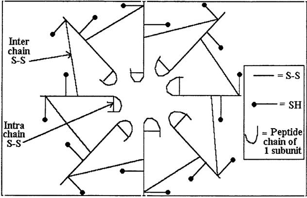
bonds must connect the subunits. One possible structure for the native (normal)
protein is shown at:
http://www.columbia.edu/cu/biology/courses/c2005/images/picture-rp3-08.jpg
B. Hint: What property (or properties) do the subunits have in common? What could be different? Consider the implications of the methods that have already been tried.
B. Answer: Column chromatography is the only method listed that is likely to work. The info given implies that the charge, molecular weight and shape of the subunits* must be the same. This follows since they act the same on starch gel electrophoresis, SDS gels and in the ultracentrifuge*, respectively. The subunits therefore should not be separable by gel electrophoresis of any kind or gel filtration. The subunits could have the same charge, molecular weight and shape and still differ in polarity and therefore solubility. So they could be separable by chromatography. It would have to be on a column since macromolecules move very slowly on paper.
*Since you did the ultracentrifugation on refolded subunits, the shapes must be the same. (You know the subunits have the same molecular weight from the SDS gel electrophoresis.) If you did the ultracentrifugation while the subunits were denatured, you wouldn't know if their shapes (when native) were the same or not. However, since you kept the protein in dilute solution and removed the mercaptoethanol and urea, the subunits could refold, but not reaggregate.
C-1 & C-2. Answer:
C-1. There are 8 subunits in the native molecule. (144,000 mol. wt total
/18,000 mol. wt per subunit = 8 subunits.)
C-2. Each subunit has 6 SH groups, so there must be 6 cysteines.
Hints for C-3 & C-4: How
many SH groups total in the native enzyme? How many are in S-S bonds? How many
S-S bonds must (or must not) connect subunits? The best way to solve this
problem is to try and draw a structure that fits the data. Use the information
given to try and figure out a plausible structure, draw a picture, and then
alter the picture as needed until it fits.
C-3 & C-4. Answer: There are 16 S-S bonds per native
molecule and one S-S bond within each polypeptide. In each native molecule,
there are 8 S-S bonds within chains and 8 S-S bonds between chains. Each subunit or chain has 2 SH groups that are
not linked up in S-S bonds, 2 SH groups linked to each other to form an
intrachain S-S bond and 2 SH groups linked to the subunits on either side. The
gentle mercaptoethanol breaks the S-S bonds between chains but not the S-S bonds
within chains. The S-S bonds within the chains are relatively resistant to
breaking by mercaptoethanol because they are buried inside the protein. The S-S
bonds between the chains are sensitive to breakage because they are exposed on
the surface of the protein.
2. A. Hint: Which amino acids have negatively charged side chains?
B. Hints:
1) Do all the sub-peptides have the same net charge at pH 7? How does this help identify any of the spots?
2) Which spot has the highest Rf? Is the most hydrophobic?
Answers to A & B.
gln-lys-ser-thr-glu-|| val-ile-phe-pro-glu- || arg-phe-leu-val-glu
A B C
Explanation: On the fingerprint, A is low middle (no net charge; didn't migrate very far -- lowest Rf), B is highest & right of center (net negative charge, and migrated farthest -- highest Rf); C is not as high, middle (no net charge, migrated an intermediate distance -- intermediate Rf. ) . Chromatography is usually done with a hydrophobic mobile phase, so highest Rf corresponds to most hydrophobic sub-peptide. In this case, the mobile phase must be hydrophobic (even if you didn't know in advance) because sub-peptide B, which is the most hydrophobic, migrates the farthest during chromatography. You know the top spot is B, because only B has a net charge at pH 7.
C. Hint: Diagram out what is being done here. Are you doing one fingerprint or two? What results will you get from the procedures described?
C. Answer: Yes. You would do two fingerprints -- one after treating a sample of the peptide with enzyme A, and one after treating a sample of the peptide with trypsin instead of enzyme A. You would then sequence each sub-peptide. You could deduce the sequence of the original peptide -- the order of the enzyme A sub-peptides -- by noting the overlap between the sub-peptides produced by the two different enzymes.