Corrections posted during the exam are included in red.
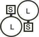 1) Protein B is a heterotetramer with 2 pairs (L
and S) of identical subunits whose subunit molecular weights are 40,000 and
10,000. The protein is shown schematically in the right, where the connecting
lines represent disulfide bonds. Indicate below the number of band(s) of
protein seen after ultracentrifugation under the indicated conditions, and the molecular
weight of the protein molecules found in that band:
1) Protein B is a heterotetramer with 2 pairs (L
and S) of identical subunits whose subunit molecular weights are 40,000 and
10,000. The protein is shown schematically in the right, where the connecting
lines represent disulfide bonds. Indicate below the number of band(s) of
protein seen after ultracentrifugation under the indicated conditions, and the molecular
weight of the protein molecules found in that band:
1A) buffer at pH7 and moderate NaCl concentration:
__1_ _________________100K_______________
40+40+10+10 = 100,
native structure intact under mild conditions
1B. as A but with 7M urea: _1__ ________________________50K___________________________
Urea disrupts all
weak bond so quaternary structure disrupted, disulfide bond keeps 40 and 10
together
1C. as B but
treated with an effective concentration of mercaptoethanol:
__2_ ____________________1 of 40K
and 1 of 10K _____________________________________
Disulfide bond now
broken and all subunits free to diplay
their individual MWs
1D. If Protein B were subjected to
SDS-polyacrylamide gel electrophoresis (no mercaptoethanol), you would expect
see __1____ band(s) of molecular
weight(s): ______________50K________________________________
As in 1B above
1E. Treatment of protein B with an effective concentration of mercaptoethanol under otherwise mild conditions followed by gel filtration results in 2 peaks of eluted protein with apparent molecular weights of 10,000 and 100,000. When the 100,000 molecule was subjected to SDS PAGE a single band of 40,000 daltons is seen. The 100,000 dalton molecules probably contain weak bonds between (circle all that apply): (L:L)(S:S)(L:S)
Weak bonds
holds the large subunits (L) together but not the small (S) subunits, which
show their monomeric MW at 10,000.
1F.Circle the factors that could contribute to these molecular weight results in1E and explain:
(shape) (friction) (ester bonds) (glycosidic bonds) (quaternary structure) (SDS) (D-amino acids)
The shape of
the 80K 2L dimer is elongated relative to a sphere, and so it appears larger (100K)
in gel filtration because it has difficulty entering channels that a sphere of
the same molecular weight could enter more easily. The association of the 2L subunits is an
example of a quaternary structure.
[ Friction affects the rate of sedimentation
in ultracentrifugation but is not a factor in gel filtration. There are no
glycosidic or ester bonds in this protein, and SDS is a detergent that is not
used in gel filtration. ] SDS effect not
sought here, its inclusion is neutral for grading.
2. D-Mannose differs from D-glucose
in having its hydroxyl at carbon 2 in the axial up position. No explanation is
required for 2A and 2B.
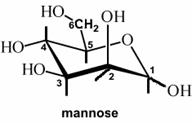 2A. In the mannose structure shown at
the right the anomeric carbon is (choose one):
2A. In the mannose structure shown at
the right the anomeric carbon is (choose one):
(0) (1) (2) (3) (4) (5) (6)
2B. In the mannose structure shown at the right, the orientation of the hydroxyl group at the anomeric carbon is (circle all correct answers) : (alpha) (beta) (axial up) (axial down) (equatorial)
2C. A polysaccharide made of repeating mannose units joined in a beta-1,4 glycosidic bonds will most likely form: (bundled straight chains like cellulose) (straight chains but unbundled) (a helix like starch) (a helix but tighter than starch) (a helix but looser than starch). Explain.
 Polymannose would not have the angled turn
to make a helix as does starch. Because the connection between all the sugars
is straight equatorial-equatorial and at opposite ends of the ring, it would
form a straight chain like cellulose and form bundles using extensive hydrogen bonding capacity of its hydroxyls.
Polymannose would not have the angled turn
to make a helix as does starch. Because the connection between all the sugars
is straight equatorial-equatorial and at opposite ends of the ring, it would
form a straight chain like cellulose and form bundles using extensive hydrogen bonding capacity of its hydroxyls.
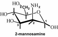
2D. In 2‑mannosamine
an amino group has replaced the hydroxyl on carbon 2 has been replaced
by an amine. You would expect a polysaccharide made of repeating 2-mannosamine
units joined in a beta-1,4 glycosidic bonds to have the physical
characteristics of:
(cellulose ) (starch) (neither) . Explain
Like polymannose, polymannosamine
would not have the angled turn to make a helix like starch. It would form a straight chain like cellulose
but would not form bundles due to the repulsion of the positively charged amino
groups.
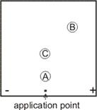
3) Suppose you subject the oligopeptide below to fingerprinting using Enzyme A, a proteolytic enzyme that hydrolyzes polypeptides after (i.e., on the carboxyl side of) amino acid residues with a negatively charged side chain. All operations are carried out at pH7. The resulting fingerprint with 3 spots is shown on the right. No explanation is required for 3A and 3B.
gln-lys-ser-thr-glu-val-ile-phe-pro-glu-arg-phe-leu-val-glu
3A. Show where Enzyme A cleaves by drawing vertical lines through the sequence.
3C. If you did not know the sequence but could sequence the sub-peptide in each spot, explain whether or not you would be able to deduce the sequence by repeating the whole experiment using trypsin, which cleaves after amino acid residues having a positive side chain?
gln-lys-ser-thr-glu-||
val-ile-phe-pro-glu- || arg-phe-leu-val-glu
A B C
[where a is low
middle (no net charge), b is highest right of center (net negative charge); c
is not as high,middle (no net charge) ]
3C. Yes, you could
deduce the sequence by noting the overlap between the sub-peptides produced by
the two different enzymes used.
4. Suppose E. coli cells contain 100 molecules of the enzyme beta-galactosidase per cell. Further suppose that the turnover number of beta-galactosidase is 200 molecules of lactose hydrolyzed to glucose and galactose per second per molecule of enzyme, and that the Km for lactose is 2 x 10⁻⁴ M. In glucose minimal medium, E. coli grows with a doubling time of 1 h at 37oC and 2 h at 30oC.
4A. If you start with 100 E. coli cells in a test tube of glucose minimal medium and incubate the tube at 30oC for 2 h and 37oC for 3 hours, how many molecules of beta-galactosidase will be present in the tube at the end of the incubation? ___________
100 à
200 at 30C; 200 à 23 = 8X à 1600 cells final
[160,000]: 1600 cells, x 100 molecules per cell à
160,000
4B. Of the number of molecules your wrote in (A) above, what percent were made during the entire incubation period? Show your calculation. ___________
[95] : 1600-100 = 1500, /1600 = 15/16 = 0.94
4C. If you break open the cells at the end of the incubation period and add 10-4 M lactose, how many molecules of galactose will be formed after 10 seconds? Be sure to show your calculation. _________
10-4M lactose = ½ Km,
so yields ~ ¼ Vmax (early ~linear part
of curve). = ¼ x 200/sec x 160,000 molecules x 10 sec.= 8 X 10⁷.
Solving using the
equation yields a more exact answer, somewhat higher.
Either way is OK.
4D. If you had incubated these bacteria for
another o hour at 37oC before
breaking open the cells and measuring the enzyme activity, the Vmax would have
been: (the same) (double) (half)
(can’t predict)
Vmax = k3Eo, so since
the number of cells would have doubled the number of enzyme molecules would
have doubled, so Eo doubled, and so Vmax would double.
5) Consider the following compounds, shown in
un-ionized form.
(I) stearic acid, a fatty acid: CH3-(CH2)16-COOH
AND
(II) phosphatidyl ethanolamine, a phospholipid.
CH2 –
|
CH –
| O
CH2- O-P-O-CH2-CH2-NH2
|
OH
5A) Which has the greater absolute net charge at pH 7? (I) (II) (same)
[I]
5B) Which is
found (even as a residue), in a cell membrane?
(I) (II) (both) (neither)
both
5C) If (I) were the only type of fatty acid in a triglyceride, ay 37oC, it would form a (solid fat) (oil) neither)
[solid fat] SInce there is no double bond to interfere
with the ready association of the hydrophobic tails by hydrophobic forces.
5D) If (I) were subjected to hydrogen gas plus platinum, then it would be expected to
(change form) (not change form) (can’t predict).
[not change form.
already saturated]