C2005/F2401 '06 -- Lecture 18
-- Last Edited: 11/13/06 05:19 PM
© Copyright 2006
Deborah Mowshowitz
and Lawrence Chasin Department of Biological Sciences Columbia University New York, NY.
Reminder: Selected problems are indicated in red bold. We suggest doing these problems as soon as possible to consolidate your understanding of the material covered in each lecture. Important Reminder: These are NOT the only problems you should do -- you should do ALL of them. For a complete list of problems corresponding to each lecture, see the problem set info page.
Handouts: 18A & 18B (Not on web). 18A = Introns & Splicing; 18B = Colony Hybridization & Probes.
I. How to find the Right Clone -- the one with the insert you want
The set up: Suppose you make a DNA library -- you carry out the procedure outlined on handout 17B, left panel. Using the techniques described last time, you cut up human DNA and make a collection of hybrid plasmids, each containing an insert of human DNA. Then you put the plasmids into bacteria, and grow up colonies of the bacteria. You now have many clones -- many colonies of bacteria. Each colony or clone contains a plasmid with a different insert. (You have already eliminated colonies of bacteria that got no plasmid or got a plasmid without an insert, using the procedures discussed last time.)
The question(s): How do you find the clone that has 'the gene of interest' -- the one you are looking for? If the gene is there, how do you find out if it is working? (You may want to be sure protein is made, or it may be sufficient just to show the DNA is present.)
An example: Let's suppose you made a human DNA library and are looking for the human gene that codes for the enzyme tryptophan synthetase. This enzyme was previously discussed as part of the trp operon in bacteria. TS = last enzyme in synthesis of trp. For the sake of this example, let's suppose that humans actually make this enzyme. (I doubt if they really do, since humans can not synthesize trp.)
What could you look for? You need to find a cell with some selectable property because it has (or is using) the gene of interest (human gene for TS, in this example). What will that property be? You can check at any step DNA (genotype) → RNA → protein → job → phenotype.
A. DNA (can be detected by hybridization) -- minimal requirements for detection -- DNA has to be there, but doesn't have to "do anything." Need not be transcribed, translated etc. You do need a probe (something to hybridize to DNA) to detect the DNA. See next section on probes.
B. RNA from DNA
(detected by hybridization with probe as
above). To make RNA requires correct transcription
signals but detection of RNA has advantages of amplification -- cell can make
many copies of RNA from the one and only copy of DNA. RNA is usually easier to find than DNA (more
copies to hybridize to probe).
C. Protein
(detected
using antibodies) -- Detection relies on structure, not function. Requires proper
processing of mRNA (not yet discussed) and correct translation signals. (But
protein need not work properly to be detected by antibodies.)
D. Job (measure function of protein -- usually enzymatic activity). Have to supply substrate. Requires all of above (transcription, translation, etc.) plus proper folding and/or modification sometimes. Note function of a protein is sometimes hard to measure if protein is not an enzyme, or requires binding to membrane, etc. in order to work
E. Phenotype (usually measure growth under certain conditions). If you start with a drug-sensitive or TS- bacterium (trp-) and add a plasmid, is bacterium now drug resistant or trp+ etc? Requires that protein be made and function well inside a cell, not just in a test tube. Added DNA must supply a new function or replace (complement) a missing or defective function. Note this one is "selectable" in terms of growth/no growth in many cases. (Replica plating on different selective media can be used to find out if clones are drug resistant, trp+ etc. Step 6 on handout 17B or 18B.)
Important: properties near the top of list require the least in terms of function, but properties near bottom are easiest to measure and/or most significant.
II. Probes
A. What's a probe? It's a nucleic acid that's complementary to the sequence you are looking for, and it is usually labeled or tagged in some way -- with radioactivity, fluorescence, or something else that's relatively easy to detect. Probe may be single stranded or double stranded.
B. Why do you need a probe? Suppose you made a human DNA library as described above. You need the probe to find the right colony (or clone) -- the colony with the gene/insert you are looking for. Probes are used in many different applications to detect DNA and/or RNA in colonies, bands on gels, etc. See below for examples.
C. How do you get a probe that is complementary to the gene of interest?
1. cDNA (works well for abundant proteins). Use reverse transcriptase to make
copy of mRNA. DNA copy of mRNA is called "cDNA."
(cDNA =
complementary DNA or copy DNA.) cDNA is commonly made and used as a
probe for a highly expressed genes -- ones that make a lot of protein and
therefore a lot of mRNA.
(DNA is more resistant to degradation and easier to work with
than RNA, which is one reason people use cDNA instead of the mRNA itself.) See Purves 16.8 (17.9).
Question: If you
have the cDNA, why do you need the gene itself? The gene or genomic DNA is
usually not the same as the cDNA,
because of introns, as explained below.
2. Oligonucleotide probes (good if know amino acid sequence) -- Sequences up to 50-100 nucleotides long can be synthesized by chemical means. An oligonucleotide of 15-20 nucleotides is usually sufficient to act as a probe. (That is, the sequence hybridizes to the gene you want and not to other genes.) You actually need a mixture of oligonucleotides -- you need more than one because the code is degenerate. Can't predict exact DNA/mRNA sequence from amino acid sequence. For example, if amino acids are lys - asp - met etc, DNA would be AAG/A CAC/T ATG etc. Don't know if base #3 is G or A; # 6 is C or T etc. So you make a mixture of oligonucleotides -- some have G at position 3, and some have A etc. (During chemical synthesis use a 50:50 mixture of G and A at position #3.) You'll get a mix of oligonucleotides, and one of the combinations will be the right one to hybridize to your gene.
3. Brute force/chance -- cut up DNA and get random collection of pieces; then use some procedure to figure out which is right piece. This sounds insane, but has been used very successfully to locate some human disease genes, as will be explained later.
D. How you use a Probe to find the 'right clone' -- Colony Hybridization -- steps 7-9 on Handout 18B - left panel (Continuation of 17B) or Becker fig. 20-30 [18-30].
Step 7. Transfer bacterial colonies to nylon or nitrocellulose filter. (Same procedure as replica plating, but colonies are transferred to a thin sheet of nylon etc., instead of to surface of a Petri dish.)
Step 8. Treat cells (colonies) to lyse bacteria, release and denature DNA. DNA remains attached to filter, but is now single stranded and available to hybridize to probe. Note you did not have to purify DNA or isolate it from individual colonies. DNA was released from each colony & denatured in situ (in place).
Note: To lyse bacteria means to break them open (by any means). In this case lysis is caused by experimental manipulations, not by a virus.
Step 9. Add labeled probe in solution; let hybridize, wash off unattached probe. (If probe is complementary to the immobilized DNA on the filter, the labeled probe will hybridize and be trapped on the filter. If probe is not complementary to DNA, it will not hybridize, and it will be washed off.)
Picture at end -- shows spot where probe hybridized. You detect location of probe by its radioactivity or other label. Now you can go back to the Petri dish shown at end of step 4 (on 17B or 18B), and isolate the colony containing the nucleic acid of interest.
III. What do you do with a clone, once you have identified it? Some possibilities.
A. Use the bacteria carrying the clone as a factory to make the corresponding protein. See Purves 16. 13 & 16.14 (17.13 & 17.14) for details and an example.
1. You use an "expression vector" = a plasmid with all the right signals for prokaryotic transcription and translation. See Purves 16.13 (17.13). (For gene therapy, you want the DNA to be "expressed" in eukaryotes, so you use a vector with eukaryotic promotors, etc.)
2. FYI: You have to clone a DNA copy of the mRNA, not the actual DNA (gene). This is because DNA has introns, as will be explained below.
B. Isolate the DNA from the clone. How do you recover the cloned DNA fragment once you have identified the right clone? Say the one for TS?
1. Lyse cells to release DNA.
2. Separate plasmid DNA from chromosomal DNA. Easy since plasmid is small and circular, unlike chromosomal DNA.
3. Treat plasmid with restriction enzyme used to make recombinant plasmid in the first place. This should release the cloned fragment. Separate fragment from rest of plasmid by electrophoresis on gels (separates DNAs by size).
C. Use clone for gene therapy.
Most cloning vectors (or vehicles) used for gene therapy are modified viruses, not plasmids. The viruses are modified versions of human viruses. Different viruses have different features -- Some are useful at targeting specific tissues, some at integrating into host DNA, some at not provoking much of an immune response, etc. Note that the added gene in some cases is designed to have a switch -- transcription of the gene and production of the protein is turned on by addition of a drug. (This is similar in principle to having an inducible operon, but the mechanism is somewhat different, because eukaryotic and prokaryotic gene regulation works somewhat differently.)
IV. Blots
A. What's a Blot? -- using probes "in situ". You can immobilize DNA on a solid support. DNA does not need to be purified first. Can be released from colonies in place (in situ) as above or "blotted" from a gel. Once DNA is stuck to a support, it can be denatured in situ. Then you can add a solution of probe (the complementary, labeled, DNA) and see if probe hybridizes, as above. You wash off unattached (unhybridized) probe and see what is left. That allows you to identify band, colony, etc. containing the nucleic acid of interest (= nucleic acid that hybridizes to and/or traps probe.) Blots allow you to test hybridization of probe to many DNA samples at once.
B. Why would you need to do a blot?
1. To find the right colony (colony hybridization). You have many colonies, and you want to find all the ones carrying a particular fragment of DNA without testing them one at a time. (Details above & on 18B.)
2. To find the right piece of DNA (as vs the right colony) -- the piece from a particular part of the genome. You have many pieces of DNA, and you want to find all the ones carrying a particular sequence. For example, to detect an RFLP (such as the one shown on handout 17A), you need to look at DNA from that particular part of the genome. But if you cut up the total DNA of an organism, you get many, many pieces. How will you find the right pieces? (Details below and on 18B.)
D. Basic procedure for detecting DNA Bands on gels Handout 18B, right panel. Italics = terms on HO. How do you find the a particular fragment of DNA, as vs. a particular colony? How do you find the piece of DNA you are looking for? See Becker, box 18C [16C] or Purves 16.3 (17.3)
1. Cut DNA up, or do PCR to amplify selected pieces.
2. Separate pieces on gel (agarose gel electrophoresis). Consider: Without probe, what would pattern of bands look like? How can you find the band you want without cutting up the whole gel into slices and testing each one? A "Southern Blot" allows you to do this.
3. Blot DNA from gel to paper or plastic (transfer to nitrocellulose.) DNA sticks to the support.
4. Denature DNA (in situ)
5. Add probe (labeled cDNA or RNA)
6. Allow DNA and probe to renature in situ. Note: You can use stringent or nonstringent conditions, depending on whether you want a only a perfect match or are looking for approximate matches too.
7. Put blot in dark next to film if label is radioactive
8. Detect bands (look at autoradiograph = developed film; use other methods if label is non-radioactive.)
E. Types of Blots (from gels) -- Terminology & Variations -- Southerns, Northerns and Westerns.
1. Southerns -- Cut up DNA, separate DNA fragments by gel electrophoresis, blot, find desired fragment by hybridization to probe.
2. Northerns --Separate RNAs by gel electrophoresis. (RNAs are small enough as is; don't cut them up first.) Then blot, hybridize as before.
3. Westerns -- Separate proteins by SDS gel electrophoresis, blot, find desired protein using antibodies specific for that protein.
F. Misc. Important Features of Blots & Probes
1. Need not purify the DNA of interest first -- you test a mix and locate where in the mix your DNA of interest is. You separate everything first by size (on gels), and then find the position of the piece you are interested in/want. Or you grow up multiple colonies containing different DNA sequences. You don't have to know in advance where the sequence you want will be. Needle in the haystack, but it works! (You spread out the hay in your haystack and glue to a support. Then cover it with magnetic particles and shake off the unattracted ones. Where there are particles, your needle is underneath. This analogy is nice but misses point that your "haystack" or "needles" are sorted by size when using gels.)
2. Can test many samples at once -- can use multiple wells and/or repeat hybridization to same blot using diff. probes.
3. Probe need not be same length as fragment -- probe can be shorter or longer than target sequence or fragment you are looking for. Probe and target need not be the same length, but there must be overlap, so some region of target hybridizes to probe. Sample must "capture" probe. (Think Velcro.)
To understand blots & probes (& review genetic engineering), wait until we explain introns and exons and then try problem 13-11, esp. parts D & E. (All the "blot & probe" problems involve introns or other complications we haven't addressed yet.)
V. Eukaryotic Gene Structure -- What do you see if you clone a gene? (Handout 18A)
A. An example of how all the cloning
procedures are used. What do
you expect if you isolate the gene for the beta chain of hemoglobin and compare
the gene to the corresponding mRNA (or cDNA)? How do you do it? See handout
18A for what is expected and what is found. (Also shown in Becker fig.
21-21 [19-21] or Purves 14.6 (14.8). )
1. Make a library of plasmids
with human DNA inserts. (Purves 16.7)
2. Identify clone with beta globin chain gene. Use mRNA or cDNA as probe to identify clone with beta globin chain gene (grown up on plasmid). Using cDNA is a common way to get a probe for a highly expressed gene -- one that makes a lot of protein.
3. Isolate DNA from your clone. Lyse cells to release DNA. Separate plasmid DNA from chromosomal DNA, and digest plasmid with restriction enzyme used to make recombinant plasmid in the first place. Separate insert from rest of plasmid by gel electrophoresis.
4. Compare mRNA and genomic DNA. Genomic DNA is longer than mRNA. So you assume DNA includes sequences on the ends of the gene that are not found in the mRNA. (Extra = spacers? regulatory regions?). If you make a hybrid with mRNA and the genomic DNA, what should the hybrid look like? You can distinguish double stranded DNA (or DNA/RNA hybrids) from single stranded DNA in the electron microscope -- double stranded regions are straighter and thicker. (See handout or texts for expected picture -- one single stranded loop.)
5. What you actually get. You get a structure with two R-loops as shown on handout 18A or Becker fig. 21-21 [19-21] or Purves 14.6 (14.8.) R loops = DNA loops formed because of RNA binding = single stranded loops of DNA formed when mRNA binds to template strand of DNA and displaces the sense strand of DNA.
6. What does picture imply? The DNA has 'extra' stretches in the middle of genes that don't show up in the mRNA. See Becker fig. 21-23 [19-23] or Purves 14.4 (14.6). What happens to the extra stretches? We now know the entire gene is transcribed, including the 'extra' stretches, and then the 'extra' stretches are spliced out of the RNA. These extra stretches are called introns or intervening sequences. Most eukaryotic genes have introns. Prokaryotic genes do not (with a few very rare exceptions which we will ignore).
B. Some implications of Splicing/Processing
1. Genetic engineering implications.
There are no introns or spliceosomes (catalysts for removing introns) in
prokaryotes. This means that genes containing introns cannot be properly "read"
in bacteria. That's why cDNA is often cloned instead of the actual gene if you
want to use the DNA to make a human protein in bacteria.
2.
Why is splicing of mRNA so rare in prokaryotes?
Messenger RNA processing of all kinds (splicing, capping etc.) is absent or
minimal in prokaryotes. Rationale? There is no separate compartment (nucleus) to
hold unprocessed stuff. In bacteria, transcription and translation occur in the
same compartment, and translation starts before transcription ends. This would
not work if mRNA had to be extensively modified before it could be translated.
(Ribosomes would attach and move down the newly made mRNA before it could be
spliced.) In eukaryotes, mRNA is processed entirely in the nucleus and then
shipped out to the cytoplasm for translation after all modifications are
finished.
3. Alternative Splicing -- allows production of multiple proteins from one gene. So the number of possible proteins greatly exceeds the number of genes. This explains why we are more complex than, say worms, although we don't have many more genes! More on alternative splicing later.
C. RNA Splicing process -- Overview. See bottom panel of hand out 18A, or Purves 14.10 (14.12) or Becker fig. 21-24 [19-24]. Additional details next time.
1. Genes consist of both introns (intervening sequences) and exons. The entire thing is transcribed, and then the introns are spliced out leaving only the exons. The unspliced RNA is known as the primary transcript. (A real primary transcript has a cap on the 5' end and a poly A tail on the 3' end as will be explained below or next time.)
Note on terminology: The "ex" in exon stands for 'region that is expressed'' not for 'region that is excised'. Region that is excised = intron. Region that ends up in mRNA (not necessarily translated) = exon.
2. Splicing occurs in several steps, all catalyzed by the spliceosome = RNP = ribonucleoprotein particle. . The spliceosome, like a ribosome, is a complex structure containing both RNA and protein. Saba Valadkhan, who was a graduate student here at Columbia in Jim Manley's lab, and a TA for this course, won the AAAS Young Scientist Award in 2004 for figuring out that some of the RNA's in the spliceosome are ribozymes -- catalysts made of RNA. See texts for details of spliceosome structure, and for which part does what, if you are interested.
3. All splicing occurs in the nucleus. After splicing is finished, what happens? The sections (introns) that were spliced out are degraded, and the mRNA containing only exons goes to the cytoplasm.
Now that you know more about introns and exons, Try problems 13-5 and 13-6, and
finish problem 13-4 (E). Be sure to do the recitation problems #10. To review genetic engineering and introns, exons, etc. try 13-7 & 13-8 .
VI. Splicing of Eukaryotic mRNA
A. What is RNA splicing? Results of R loop experiments discussed above indicate that eukaryotic genes contain sections that are represented in the mRNA (exons) and sections that are not (introns). For example, you can have a gene with sections A-B-C that produces an mRNA containing sections corresponding to only A and C. (Each letter represents a stretch of nucleotides. See handout 18A. ) How is this done? The entire DNA is transcribed and then the section corresponding to B is removed. This process is known as RNA splicing, as explained above.
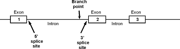
B. A typical picture of a gene with introns and exons. The picture below shows a section of the sense strand of the DNA that includes a gene with 3 exons and 2 introns. (The picture on the handout has 2 exons and one intron.) Conventions:
The picture on the handout shows double stranded DNA, but genes are often shown as in the picture below, with only the sense strand actually drawn in.
Transcription would start at the 5' (left) end of exon 1 and go to the right.
Important features of intron: Branch point, 5' splice site (also called the donor site) and 3' splice site (also called acceptor site). These are shown for the first intron only. (See also fig. 21-23 [19-23] in Becker or 14.10 (14.12) in Purves.)
Also note that the region to the left of exon 1 is NOT an intron -- it is not part of the gene. It is part of a spacer in between this gene and the previous one.
C.
Splicing Details -- See bottom of handout 18A.
1. General Features
a. Splicing out of each intron occurs in 3 steps (see handout 18A at the bottom, steps 7-9). At each step the parts of the transcript are held in place by the spliceosome. The steps are repeated for splicing of each intron -- many RNA's have many introns. (The hemoglobin beta chain transcript actually has 2.) Details are below.
b. The splice junction at the 5' end of an intron is called the 5' or donor site; the splice junction at the 3' end of an intron is called the 3' or acceptor site.
2. Steps of splicing. See handout 18A at bottom. See also Becker fig. 21-24 [19-24] or Purves 14.10 (14.12). All the steps are catalyzed by the spliceosome. Steps on handout are as follows:
(1 to 6) = transcription (and addition of 'cap' and poly-A 'tail') -- these steps to be discussed next time.
(7) RNA transcript forms loop for removal of intron.
(8). 5' splice site (donor site) is cleaved & loose end of intron (5' end of intron) attaches to branch point in the middle of the intron, forming lariat-shaped structure.
(9). The 5' donor site attaches to the 3' acceptor site, joining the two exons and releasing the intron in the form of lariat. The lariat will be degraded and the nucleotides will be recycled. The RNA containing the exons (without the introns) will be transported to the cytoplasm and translated.
3. N.B: Prokaryotes do not have introns and lack the enzymes to remove them.
D. Do exons and translated regions coincide? See diagram at bottom of 18A. Note that the start and stop signals for translation are not at the ends of the exons -- the exons include untranslated regions (5' UTR's and 3' UTR's or leaders and trailers). Therefore the exons are not = protein coding sequences, as some texts imply. (The diagram in Purves 14.4 (14.6) is incorrect.) Exons = sections of genes that are represented in the mRNA. Exons include untranslated 5' and 3' regions as well as the translated regions.
1. Leaders. At the 5' end, there is a 5' untranslated region (UTR) or
leader before translation begins (before the first AUG) that is part of the
first exon.
2. Trailers. At the 3' end, there is a 3' UTR or trailer that is after
the stop codon that is part of the last exon.
Is an exon a "coding region"? It is a "coding region" in the sense that it codes for mRNA, but NOT in the sense that it (necessarily) codes for protein. Exons contain sequences that are represented in the mRNA but do not code for amino acids.
Next time: We'll take a closer look at steps 1-6 -- how the RNA is made and gets a 'cap' and poly-A 'tail'. Then we'll do the mitotic chromosome cycle, and the actual steps of mitosis; then meiosis and some implications.
© Copyright 2006 Deborah Mowshowitz and Lawrence Chasin Department of Biological Sciences Columbia University New York, NY.