C2006/F2402 '04 OUTLINE OF LECTURE #14
(c) 2004 Dr. Deborah Mowshowitz, Columbia University, New York, NY. Last update 03/10/2004 10:43 AM . A few details and references to problems added after lecture; significant changes are in
this color.Handouts: Need 13B (Hormones Overall), 14A (Thyroid, catecholamines),14B (glands, organs), 14C (Homeostasis), 14D (Temperature Regulation)
I. Details of HT/Ant. Pit. Axis, cont.
A. Hypothalamic Hormones
1. Inputs: Neuroendocrine cells in HT produce hormones -- in response to multiple inputs -- see last lecture.
2. Outputs: Neuroendocrine cells in HT are two kinds:
a. Some have bodies in HT and axons/terminals in posterior pituitary -- release ADH & Oxytocin. See last lecture for details.
b. Some cells in HT release hormones from HT itself.
(1). Release hormones into portal vessel (connects two capillary beds) that goes direct to anterior pituitary. See Purves 41.7 [38.5] and handout 13B.
(2). Hormones released by HT affect production/release of other hormones by ant. pit.
(3). Affect on release can be stimulatory (RH's such as ACTH-releasing hormone) or inhibitory (IH's such as prolactin release-inhibiting hormone = PIH) For a complete list see Purves 41.2 [table 38.2]
(4). All HT hormones (except PIH) are peptides/proteins.
(5). PIH (prolactin inhibiting hormone) = dopamine = modified amino acid. Additional info on dopamine (DA) & related compounds -- see (a) to (d) below:
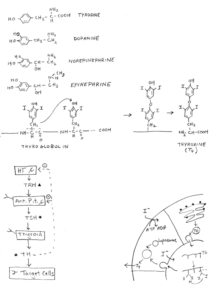
(a). Structures: See handout 14A for structures of catecholamines = epinephrine aka adrenaline, norepinephrine aka noradrenaline, and dopamine. These are all modified amino acids derived from tyrosine. All water soluble. (Note thyroxine is also derived from tyrosine but is lipid soluble; see below.)
(b). There are multiple receptors for all of the catecholamines. Receptors for epi and nor epi are called adrenergic receptors. Dopamine has its own receptors.
(c). All receptors for all catecholamines are G protein linked; effects of hormones on any particular cell type depend on (i) what receptors are present and (ii) what G protein each receptor activates. Each G protein does one of the following: activate adenyl cyclase; inhibit adenyl cyclase; activate phospholipase C. See Lecture 12 for examples of different responses to epi due to diff. receptors.
(d). All adrenergic receptors bind to both epi and norepi. Some receptor types bind better to one, some to the other, some equally well to both. Epinephrine acts mostly through beta adrenergic receptors; norepinephrine mostly through alpha adrenergic receptors.
B. Hormones of Anterior Pituitary
1. Tropic Hormones (for names of hormones and target cells see handout 13B)
a. Made by ant. pit and influence other endocrine glands
b. Release controlled by hormones from HT
c. Effect on target tissue
(1). Effect is usually release of another hormone
(2). Hormones released by targets are steroids or act like them (thyroxine)
(3). All tropic hormones work through G linked receptors and cAMP.
d. Three major tropic hormone types -- each type named after its target -- see handout 13B & table below.
See problem 7-4. (Skip 5 for now.)
2. Other Hormones of ant. pit.
a. GH and prolactin -- "pseudo tropic" hormones
(1). Similar in structure to each other (homologous) and use a special type of TK receptor
(2). Stimulate production of secretions, but not from endocrine glands.
(a). GH stimulates liver (& possibly other tissues) to produce insulin-like growth factors (ILGF 1 & 2); ILGF's from liver released into blood (act as endocrines); ILGF's from other tissues act as paracrines. (GH has other effects as well.)
(b). Prolactin stimulates mammary (exocrine) gland to produce milk. (Need oxytocin to eject the milk.)
(c). What's the difference between endocrine & exocrine glands? See handout 14B.
(i). Exocrine Gland
When gland forms, epithelial layer leaves duct to outside.
Secretion from gland flows into duct --> outside or lumen.
Examples:
sweat, mammary & tear glands --> outside; stomach glands --> lumen.
(ii). Endocrine Gland
When gland forms, epithelial layer pinches off leaving no duct
Secretion (hormone) from gland enters blood.
Example: gonads, pancreas, adrenal.
Try problems 7-1 & 7-13.
b. MSH etc.
(1). All come from cleavage of single peptide precursor (pro-opio-melanocortin or pomC) that is cut up to give ACTH and MSH etc.
(2). Same precursor can be cut up different ways in different tissues and/or species. Note: this is alternative processing of a protein, not an RNA.
(3). Function of these hormones relatively obscure.
3. Summary of Tropic & "Pseudo-Tropic" Hormones of Ant. Pit
|
Tropic (or Pseudo-Tropic) Hormone(s) |
Target Organ |
Hormones/Secretions Made by Target Organ |
|
ACTH (adrenal cortex tropic hormone) or adrenocorticotropin |
Adrenal Cortex |
Glucocorticoids, Mineralocorticoids & sex steroids* |
|
Gonadotropins -- LH and FSH |
Gonads |
Estrogens, androgens & progesterone* |
|
TSH (thyroid stimulating hormone) or Thyrotropin |
Thyroid |
Thyroxine* |
|
GH (growth Hormone) or somatotropin |
Liver (& others) |
Insulin-Like Growth Factors (ILGF 1 & 2) or somatomedins |
|
Prolactin |
Mammary Gland |
Milk |
* All lipid soluble hormones travel through the blood attached to plasma proteins.
Try Problem 7-2 & 7-4 if not yet done, but skip 5 (of 7-4) for now.
C. HT/Anterior Pituitary Axis -- Set up & Regulation of overall circuit (HT --> Ant. Pit. --> target)
1. General case: See Purves 41.8
a. The cascade: HT --> releasing hormone --> AP --> tropic hormone --> TARGET GLAND --> hormone --> TARGET TISSUE --> action.
b. Regulation: Hormone (thyroxine, sex steroids, etc.) has negative feedback effect on HT and AP. Why inhibit both?
(1). Feedback inhibits HT -- alters production of releasing/inhibiting hormones (changing signal to AP)
(2). Feedback inhibits AP response to signal from HT (probably by down regulating receptors)
(3). Both neg. feedback effects lead to inhibition of release of tropic hormones from AP
2. Specific case: thyroxine production (See handout 14A; Purves fig. 41.9 [38.8])
a. The cascade: HT --> TRH --> AP --> TSH --> TARGET GLAND --> TH --> TARGET TISSUE --> increase in BMR, etc.
b. Regulation
(1). Negative Feedback: TH inhibits production of both TSH and TRH.
(2). Two different types of goiter (enlarged thyroid)
(a). When TH is low (hypothyroidism): Lack of iodine or other factor --> low level of TH --> lack of negative feedback to HT &/or AP --> overproduction of TSH --> goiter
(b). When TH is high (hyperthroidism): Can sometimes still have too much stimulation of TSH receptors due to overproduction of TRH and/or TSH or overstimulation of receptors in spite of high TH levels (& neg. feedback). See Graves disease below.
(3). Graves disease = antibodies to TSH receptors act as agonists of TSH. (Case of (b) above). Two important terms:
agonist = acts like -- or has same effect as -- normal ligand
antagonist = blocks action of -- or effect of -- normal ligand
(4). What does thyroxine do? Raises BMR and is needed during childhood for brain development.
(5). How is thyroxine made? By modification and rearrangement of tyrosines in thyroglobulin (TG) -- see handout 14
protein (TG) made on RER --> Golgi --> vesicles
TG stored in lumen of gland
I- taken up into gland; I added to tyrosines of TG in lumen; one modified tyrosine added to OH of another.
Degradation of TG in lysosomes --> releases T4 or T3 --> diffuses out of cell across membrane. Acts like a steroid. (For structures see handout or texts.)
(6). How does thyroxine travel through the blood? All lipid soluble hormones are attached to plasma proteins, either to general proteins or specific binding proteins for that hormone. T4 and T3 are transported by thyroxine-binding globulin, which is specific for thyroxine.
Try problem 7-5 & 7-9. (If you have time, there are additional problems on this topic -- most of problem set 7. )
How do you use hormones to control homeostasis?
II. Introduction to Physiology & Multicellular organisms
A. Single cell Life Style vs. Multicellular
1. Single celled organisms
a. Surrounded by external environment -- Can't change or regulate it
b. Have one basic function -- grow and multiply
c. Respond to external conditions (since can't change them) to maintain optimal intracellular state
(1). Pick up and/or dump what is necessary for metabolism
(2). Keep intracellular conditions (pH, level of amino acids, oxygen, etc.) as constant as possible and expend minimal energy by adjusting rates of transcription, enzyme activity, etc.
d. Note no specialization: each cell does all possible functions
2. Multicellular organisms & Homeostasis
a. Each cell in organism surrounded by internal environment. Extracellular fluid that makes up internal environment is composed of:
plasma = liquid part of blood = fluid between blood cells
interstitial fluid = fluid between all other cells
b. Organism as whole can regulate composition of internal environment (milieu); therefore can maintain relatively constant external environment for each cell. Process of maintaining a relatively constant internal environment (of whole organism) = homeostasis.
c. Each cell has two basic functions
(1). Grow or maintain itself as above
(2). Specialized role in maintaining homeostasis of whole organism
d. Cells are Specialized. Maintenance of homeostasis requires co-operation of many different cell types, not just circuits within a single cell.
B. Organization -- How are cells set up to co-operate in a multicellular organism?
1. Cells, Tissues & the 4 major tissue types -- see lecture #4, & Purves fig. 40.2, 40.3, & 40.4.
2. Organs
a. Made of (different kinds of) tissues.
b. Example: lining of GI tract. Has layers -- epithelial, connective, muscle, and nervous for absorption, support, contraction, and regulation respectively. (see Purves fig. 40.2 [37.2])
3. Systems -- Group of Organs --> body or organ system. Work together to maintain homeostasis for some component. Number of systems depends on who's counting. Usual # is 8-12; see Purves for a list and illustrations (Table 40.1 [figs. 37.3 to 37.8])
III. How is a component of the internal milieu regulated?
A. Let's look at a specific example, namely blood glucose. The see-saw view. See handout 14C. or Purves 50.20 [47.19]
1. Have a regulated variable -- glucose level in blood
2. Need a sensor (or receptor) -- to measure levels of regulated variable (glucose)
3. Need effector(s) -- to control levels of regulated variable (glucose) -- usually have two effectors that act in opposing ways.
Note: The term "effector" is used differently in molecular biology and in physiology. In physiology, it is usually used to mean "a tissue or organ (like muscle or liver) that carries out an action and thus produces an effect." In this example, the effectors = structures that act to raise or lower the blood glucose. In molecular biology, the term "effector" is usually used to mean "a modulator of protein function." A modulator = a small molecule (like an inducer, enzyme activator etc.) that binds to a protein, alters the shape and/or function of the protein, and thus triggers an effect.
4. Have a set point -- the level the regulated variable (blood glucose) should be. Set point is also some times used to mean the level at which corrections (to raise or lower the value) kick in.
In most cases, there is no difference between these two definitions of set point. In some cases, the desired value (first definition) and the value at which corrections occur (second definition) may be different. For example, there may be two cut-off points-- upper and lower, that bracket the desired level of a regulated variable. At levels above or below the respective cut-off points, messages are sent to the appropriate effectors to take corrective action. The term "critical values" is sometimes used instead of "set points" to describe the cut-off point(s).
5. Negative Feedback -- system responds to negate changes from set point. (In positive f.b., system responds to change by making it bigger and bigger until --> boom!)
6. Value of regulated variable does not remain exactly constant, but stays within narrow limits.
See problem 5-1 & 5-2 a & b.
Actual Class Lecture ended here. The remaining material will be covered in Lecture #15.
B. Another example -- regulation of body temperature (in humans) -- the see-saw view (14C)
1. Features not found in glucose case:
a. Multiple sensors in different places (for core and skin temp.)
b. Need separate integrative center (IC).
(1). In this example, IC = hypothalamus (HT)
(2). Separate IC needed if there are multiple sensors.
(3). Role of IC: Co-ordinates incoming (afferent) information from sensors; sends outgoing (efferent) information to effectors.
2. Different body systems involved as effectors
|
Effector |
Action To Raise Temp |
Action To Lower Temp |
|
Skeletal muscles |
Contraction generates heat (shivering) |
None |
|
Smooth muscle of peripheral blood vessels in skin |
Muscles contract; vessels constrict to reduce heat loss |
Muscles relax; vessels dilate to increase heat loss |
|
Sweat glands |
None |
Produce sweat; evaporation increases heat loss |
|
Brain |
Behavioral (nonphysiological) responses-- put on coat, curl up, etc. |
Behavioral (nonphysiological) responses -- take off coat, etc. |
3. Cooling vs. Heating -- What can effectors do? Effectors can increase or decrease heat loss; can only increase heat generation. (No air conditioner.) Therefore ability to cool down is less than ability to heat up.
4. Metabolic rate and temperature control in homeotherms (organisms with aprox. const. internal temp.) that are endotherms (generate own heat internally) as vs. poikilotherms/ectotherms. (See Purves p. 700 [p. 817] for further discussion of these terms.) See handout 14D, top.
a. Constriction/dilation of blood vessels uses relatively little energy. This allows adjustment of body temperature without changing metabolic rate in range of external temperature called the "neutral zone."
b. Both heating (by shivering) and cooling (by sweating) require lots of energy. Therefore MR (metabolic rate) increases outside neutral zone at both high and low temperatures.
c. Overall how MR (metabolic rate) changes with external temperature (see handout 14D, top or Purves, fig. 40.15 [37-19]). Thermo-neutral zone is bounded by critical temperatures = points at which shivering or sweating occur = set points for shivering or sweating.
C. Body Temperature and the General Case -- The Circuit View -- see handout 14D, bottom.
1. Signals: System involves more than one cell so signals must be sent using nerves and/or hormones from sensor to effectors. (For comparison: only 1 cell involved in 2 cases of f.b. previously discussed: Fe circuit with Ferritin/Transferrin Receptor or interconversion of glycogen and glucose 1P in skeletal muscle)
2. Afferent vs Efferent Signals. Bottom half of circuit has two arms -- Afferent information (from sensors in to IC) vs efferent (out of IC --> effectors)
3. Regulation vs Control. Note variable (glucose level) is said to be "regulated" but processes that alter levels of regulated variable (glucose uptake, release, etc.) are said to be "controlled."
4. May be multiple effectors and/or sensors.
5. IC = nervous tissue or brain.
a. Compares current value to set point; sends appropriate message to effectors.
b. Can adjust set points and/or critical points. Fevers & feedforward.:
(1). Fevers -- Raise set point(s)/critical points for shivering/sweating
Shift curve of MR vs external temp to right; shivering and sweating start at higher temps. (so don't have to cool off as much to start shivering and need to heat up more to stop sweating); raises body temp. above normal. Raises set point (desired level) of internal body temperature.
Why fevers? High temperature interferes with bacterial iron metabolism & improves immune function.
(2). Feedforward or anticipation -- altering set points and/or critical points to adjust to anticipated factors. Planning ahead. Examples:
Body temperature: Skin temperature affects critical temperature/set points for generating heat and/or shivering. If body is cold, but it's warm outside, shivering can be postponed, saving energy, and you'll still warm up. This = lowering set point/critical points for shivering, not changing set point of internal body temperature. Changes what effectors you use to warm up, not end result. (See Purves, fig. 40. 19 [37.23]).
Secreting insulin when you start to digest food in the stomach, but before the digestion products (glucose, amino acids etc.) reach the blood. This way tissues will be ready to take up the glucose as soon as it enters the blood.
D. What other components of internal milieu are regulated besides glucose, temperature? Many nutrients like amino acids; concentrations of water, salts and ions (Na+, K+ etc.), gases (CO2, O2), waste products, volume & pressure of blood, and pH.
IV. Matching circuits and signaling -- an example: How the glucose circuit works at molecular/signaling level
A. Re-consider the circuit diagram for homeostatic control of blood glucose levels -- what goes along the arrows, and what happens in the black boxes? (See handouts 14C & 14D or Purves 50.20 [47.19])
B. Mechanism of Action of hormones Involved
1. Insulin
a. Receptor: Insulin works through a special type of tyrosine kinase linked receptor; See Purves 15.7. Insulin has many affects on cells and the mechanism of signal transduction is complex (activating multiple pathways). In many ways, insulin acts like a GF (it has GF like effects on other cells).
b. Effect on GLUT 4: In some tissues (muscle, adipose), insulin mobilizes transporter for facilitated diffusion (of glucose) -- GLUT 4 protein -- promotes fusion of vesicles containing the transporters with plasma membrane.
c. Other Effects: In other tissues, insulin promotes utilization of glucose.
d. Overall: promotes uptake & utilization of glucose.
2. Glucagon
a. Receptor: Glucagon works through a G protein linked receptor that triggers the cAMP pathway (as for epinephrine).
b. Effects: Effects on tissues vary; generally promotes production/release of glucose, not uptake or utilization.
c. Uses same pathway as epinephrine. Note that same pathway can be used for two different hormones (epinephrine & glucagon)
(1). Both receptors trigger same pathway --> cAMP --> etc. so get same response to both hormones in same tissue.
(2). Receptors present determine which tissues will respond to each hormone. Muscle has Epi receptors and responds to Epi but not glucagon; liver has receptors for both and responds to both.
(3). Two hormones control same process (glycogen metabolism) for different ends -- Epi controls response to stress; glucagon response to low blood sugar (homeostasis).
(4). Same hormones give different response in liver vs adipose. How? Different enzymes and processes (glycogen metabolism vs. fat metabolism) available to be controlled.
Next Time: More detailed look at the glucose circuit -- the Absorptive State and the Post-Absorptive State.