C2006/F2402 '04 Lecture #22 -- Gas Exchange cont.; Kidney Function; Salt & Water Balance
(c) 2004 Deborah Mowshowitz . Last updated 04/20/2004 01:07 PM.
Handouts: 21C (from last time = Purves 48.17 & 48.12, 4-6)); 22A -- Regulation of Gas Exchange; 22B Kidney Structure; 22C Kidney Processes (= Purves 51.7) & Tubular Cell Structure (like Purves 51.6)
I. Gas Exchange -- how do you get O2 to cells and CO2 to lungs? See Gas Exchange handout (21C) & Purves 48.18 (45.17).
A. Exchange (of oxygen, carbon dioxide & other nutrients and waste) at cellular level -- what happens at the capillaries. (For structures see Purves fig.48-12 ( 45-12) or handout 21C.
1. In lungs: Materials in alveoli exchange with materials in pulmonary capillaries
2. In tissues: Materials in cells exchange with materials in systemic capillaries
3. Structure of capillaries: Capillaries have large surface area, thin walls, and slow flow of blood, promoting exchange
4. Structure of alveoli: Alveoli (sacs containing air) have huge surface area, thin walls, and are very close to capillaries, promoting exchange.
B. How is O2 carried?
1. Lungs: Hemoglobin (Hb) binds & traps O2 inside red blood cell (RBC) at lungs (See handout 21C, panel B). Very little oxygen is dissolved in plasma.
2. Tissues: Hb releases O2 in tissues (See handout 21C, panel D)
3. Important properties of Hb that enable it to function properly -- see curve of % saturation of Hb vs pO2. Purves fig. 48.14 (45.14). Note sigmoid shape of curve. pO2 = partial pressure of O2 = measure of concentration of free (not bound) O2 = measure of concentration of O2 dissolved in blood or liquid part of tissue.
a. Plateau means Hb always saturated with O2 in lungs.
b. Curve is steep in range of pO2 found in tissues -- so releases O2 as needed.
c. Sigmoid curve indicates allosteric enzyme; binding of O2 is co-operative. O2 affinity shifts with amount of O2 already bound/lost. Promotes tendency to empty of fill up completely (with O2).
d. Affinity of Hb for O2 can change due to genetic [Purves fig. 48.15 (45-15)] or environmental differences [Purves fig. 48-16 (45-16)].
(1). Lower affinity shifts curve to right; higher affinity shifts curve to left (takes less O2 to fill Hb up).
(2). Ligands bound to Hb can change affinity and shift curve. For example, DPG (2,3 diphosphoglycerate) -- found only in RBC -- shifts curve to right ---> optimal affinity for job
(3). Changing amino acid sequence slightly can shift curve right or left. Example: HbF (fetal Hb) has higher affinity; suits its job. See Purves fig. 48.15 (45-15)
(4). Conditions in tissues (Low pH, high CO2) shift curve to right, allowing Hb to unload O2 where needed, and H+ and CO2 to bind to deoxygenated Hb. (Reverse happens in lungs.) See Purves fig. 48-16 (45-16).
Try problems 10-7 & 10-13; then try 10-14 & 10-16.
C. How is CO2 carried? See handout 21C panels A & C or Purves fig. 48.17 (45.17)
1. What happens in tissues? (panel C on 21-C)
a. CO2 from metabolism enters RBC; relatively little is dissolved in plasma.
b. Inside RBC, carbonic anhydrase (one of fastest enzymes known; turn over # of 6 X 10⁵/sec) converts CO2 to carbonic acid. Traps CO2.
c. Carbonic acid disassociates into bicarbonate and H+
d. Anion exchanger (band 3 protein) switches bicarb (in cell) for Cl- in blood.
e. Some H+ & CO2 binds to Hb (deoxygenated)
2. What happens in lungs? (Panel A on 21-C)
a. Process described above reverses -- bicarb. reenters cell, made back into CO2, etc.
b. CO2 released to air (low CO2 in alveloli/air pull CO2 off by Le Chatelier's principle; higher CO2 conc. in blood than in alveoli)
c. O2 helps drive off CO2 and H+ from Hb. (Additional push factor.)
II. Regulation of Gas Exchange
A. Acid problem
1. Acidity & CO2: Whenever CO2 is too high, system gets too acid and vice versa. (Because of carbonic anhydrase rxn. etc. See above.)
2. Breathing rate & acidity: Breathing too slowly --> acidosis. (Loss of CO2 is too low.) Therefore control of pH done by controlling breathing rate.
B. How are O2 and CO2 levels regulated? What processes are controlled?
1. Control of blood flow in tissue capillaries -- more flow gives more exchange. See Handout 22A and Purves fig. 49.18 [46.19]
a. Local control of smooth muscle by O2 and CO2 -- In tissues, high CO2, low O2 promote relaxation of smooth muscle in arterioles feeding the capillary beds -- favors higher blood flow. (This is not the case in the lungs. See key to RP10.)
b. Hormones and Autonomic system can affect flow too. (Effect depends on transmitter and/or hormone and type of receptors.)
2. Control of breathing rate -- breathing faster moves air faster and fosters exchange in lungs. Details below.
3. Control blood pressure overall (thereby affecting flow) -- to be discussed later after water/salt balance.
C. Mostly CO2, not O2 levels regulated.
1. Sensors for CO2 levels more sensitive than for O2 levels.
2. CO2 level is major indicator -- See Purves 48.19 [45.19].
a. Can consider level of CO2 as indicator of state of gas exchange -- indicates
(1). Waste product levels
(2). pH
(3). Metabolic demand -- if CO2 high, means metabolism is running quickly and need more O2. (So don't need to measure/correct O2 levels separately.)
b. Max. levels of total or available O2 (in oxygenated blood where sensors are) don't change much because of plateau in Hb O2 binding curve. pO2 in deoxygenated blood does change; it depends on how much O2 is taken up by the tissues.
D. Regulation of Breathing (see Handout 22A)
1. Medulla = IC; generates own rhythm (has pacemaker cells but rhythm modified by other factors). See Purves 48.18 [45.18]
2. Inspiratory Neurons control Primary effectors = muscles of inspiration (inhalation)
a. Inspiratory (inhalation) neurons --> motor neurons --> contraction of inhalation muscles = external intercostals (between ribs), diaphragm. (Insp. neurons are not themselves motor neurons.)
b. Relaxation (expiration) is passive unless demand is high
c. If necessary, expiratory neurons trigger contraction of expiratory (exhalation) muscles = internal intercostals, abdominals
3. Regulated Variables = Levels of CO2, O2
4. Sensors -- see Purves 48.20 [45.20]
a. Peripheral chemosensors -- In major arteries (respond primarily to low O2; less sensitive to changes in pH and CO2.)
b. Central chemosensors -- medulla (primary CO2 sensor -- actually senses mostly H+)
c. Stretch sensor in lung -- responds to stretch; gives slowdown override signal in extreme cases if breathing too fast (Hering-Breuer reflex)
Try problems 10-8 & 10-10.
III. Kidney Structure & Function (Handout 22B). See also Purves figs. 51.7, 51.8, 51.10, 51.11 [48.7, 48.8, 48.10]
A. Overall Function -- what does the kidney do?
1. Function: Controls water loss and determines what other specific components will be excreted and what will be retained.
2. How does it carry out its function? Carries out filtration, tubular secretion, & tubular reabsorption & then controls volume. See handout 22-C or Purves 51.7.
a. Concentration/osmolarity: Urine can be more concentrated than the plasma (but can vary concentration and/or volume to suit need) -- controls volume of body fluids -- plasma, extra cellular fluid, etc. (blood pressure)
b. Result of tubular (selective) secretion: Carries high concentrations of certain dissolved materials (secreted by cells lining the lumen) -- removes waste, toxins
c. Result of tubular (selective) reabsorption: Does NOT carry other materials (which are selectively reabsorbed) -- conserves valuable materials
Try problem 12-3.
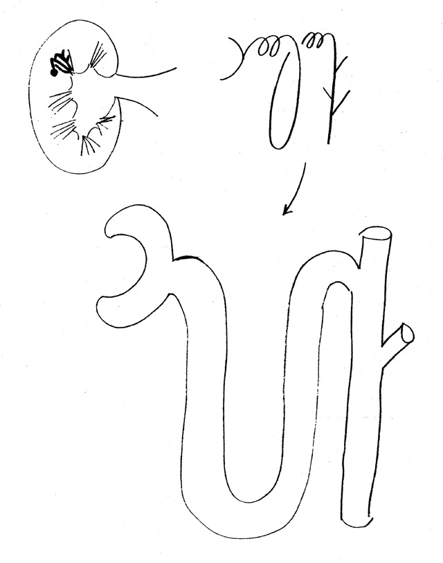
B. Overall structure -- see handout 22-B or Purves fig. 51.10 & 51.11 [48.10]
1. Kidney has medulla (inner part) and cortex (outer)
2. Functional unit = nephron (Purves 51.7 [48.7])
3. Visible unit (in medulla) = Renal Pyramid = bottoms of many nephrons
4. Tops of nephrons in cortex
C. Structure of Nephron -- see handout 22-B & C or Purves fig. 51.7 & 51.11 [48.7 & 48.10]. For EM pictures see Purves 51.8.
1. Nephron itself -- parts in cortex
a. Bowman's capsule
b. proximal (convoluted) tubule
c. distal (convuluted) tubule2. In medulla
a. Loop of Henle
b. Collecting duct (shared by many nephrons)3. Capillaries
a. 2 sets in series
(1). Glomerular
(a). form glomerulus inside Bowman's capsule
(b). function = filtration(2). Peritubular
(a). surround tubules
(b). vasa recta = part in medulla
(c). function in secretion (concentration of substances in filtrate) & reabsorption (removal of substances from filtrate)
Note: Cells lining tubule do actual secretion/reabsorption but capillaries remove reabsorbed material or provide material to be secreted. (see below)
b. How capillaries connected
Afferent arteriole --> glomerular capillaries ---> efferent arteriole --> peritubular capillaries --> venule
D. Fine structure/Function of Nephron -- Let's follow some liquid through -- any details not covered in remainder of this lecture will be covered next time.
1. Filtration in glomerulus
a. About 20% of blood liquid (plasma) enters Bowman's capsule = filtrate
b. Filtrate contains no large proteins or cells
See problem 12-6.
2. Structure of cells lining tubules -- see handout 22C or Purves fig. 51.16, for an example [or see 48.10])
a. Tubules lined by layer of polarized epithelial cells (similar to those lining intestine)
b. Epithelial cells have different proteins/channels/transporters on apical or luminal surface (facing lumen) and baso-lateral surface (facing interstitial fluid and capillaries)
c. Cells in different parts of tubule have different transporters/channels on apical surface
d. Materials must cross epithelial cells to enter or exit lumen of tubules.
e. Depending on proteins in apical surface, cells secrete materials into lumen or reabsorb material from lumen.
f. Interstitial fluid separates epithelial cells and capillaries.
3. Reabsorption in proximal tubule
a. Many substances removed from lumen by secondary act. transport
(1). examples: glucose and amino acids
(2). Cross apical surface of epithelial cell by Na+ cotransport
(3). Exit baso-lateral side of cells into intersit. fluid by facilitated diffusion
(4). Process is similar to absorption in cells lining intestine
b. Na+/K+ pump on basolateral side keeps internal Na+ low.
c. Water follows salt.
4. Events in Loop of Henley and rest of tubules -- overall picture of state of filtrate
a. Events in Loop
(1). Osmolarity (solute concentration = concentration of dissolved material) increases as descend due to loss of water
(2). Osmolarity decreases as ascend due to loss of salt; reaches min. value less than that of blood. Therefore can excrete urine that is hypo-osmotic (less concentrated) than blood.
(3). Net effect of going through countercurrent loop -- less volume, less total salt to excrete (even if filtrate and blood are iso-osmotic when done).
b. Events in distal convoluted tubule (& first part of coll. ducts) depend on aldosterone -- promotes pumping of Na+ and water follows.
c. Events in collecting duct depend on ADH -- Osmolarity will increase (and volume decrease) in collecting duct if ADH (vasopressin) present and water removed
5. Details of Loop of Henley (See Purves 51.12 [48.11])
a. Cells in descending loop and lower part of ascending loop are permeable to water
b. Cells in rest of ascending are impermeable to water and pump NaCl out from lumen to interstitial fluid
c. NaCl removed from ascending loop accumulates in medulla, forming a gradient of increasing osmolarity (salt concentration) as reach bottom of loop = core of medulla. (Multiplier effect)
d. Filtrate from proximal tubule loses water as it descends into medulla --> high concentration NaCl --> to be removed in ascending
e. If NaCl diffuses into descending loop, it is carried around and pumped out in ascending = escalator effect.
f. Why called countercurrent? Because flow in two sides of loop is in opposite directions physically and with respect to osmolarity. First leg (descending) of loop removes water --> higher osmolarity; second leg (ascending) removes salt --> lower osmolarity.
See problems 12-1 & 12-2.
6. Distal Tubule and Collecting Ducts
a. Filtrate entering here is at minimum osmolarity
b. More water and/or Na+ removed under influence of aldosterone and ADH
c. Role/Mech. of action of ADH
(1). ADH (using cAMP) stimulates insertion of water channels/pores into membranes of collecting duct (and maybe late distal tubule)
(2). Water flows out ADH-stimulated channels (if in membrane) because of salt gradient in medulla.
(3). Diabetes insipidus -- result of no ADH or no response to ADH
d. Role of aldosterone
(1). Promotes reabsorption of Na+; water follows.
(2). Amount of Na+ reabsorbed due to aldosterone is small % of total, but adds up.
See problem 12-11.
Next Time: Wrap up of kidney function, Regulation of kidney function & Summary of all circuits affecting blood pressure; then on to immunology.