C2006/F2402 '04 -- Outline on Immunology -- revised 4/25/03
(c) 200
4 Dr. Deborah Mowshowitz Columbia University, New York, NY. Last Update: 04/27/04 07:43 PM . The order of topics in the lecture and the order of topics in the problems do not match, so you may find it easier to do all the problems after reviewing the lecture. By the end, you should be able to do problems 13-4 to 13-12.| Cells | Secreted Proteins | Cell Surface Proteins |
| B cells | Antibodies (Ab or immunoglobulins; 5 classes) | MHC |
| TC cells | Perforin | BCR |
| TH cells | Cytokines (Interleukins & interferons) | TCR |
| phagocytic cells | CD4 | |
| APC's | CD8 |
The chart above summarizes the major players in immunology. By the end of the next lecture, you should be able to describe what each item is, its significance, and how it is related to all the others.
Handout: 224A (Antigen Presenting Cells & Activation of T
cells) -- Posted version is from a previous year; there are some minor
differences.
Handout 24B is not on web; it includes clonal selection (Purves 19.7), T & APC
cell interactions (like Purves 19.17) & activation of B cells (like Purves 19.18
(a) -- to be discussed next time).
I. Specific (or Acquired) Immune Response -- Major Features
A. Intro. What are major components of the specific immune system?
1. Proteins -- antibodies, TCR's & MHC's. See lecture 23.
2. What Cells are involved? (See bottom of 23B.) White blood cells (leukocytes) -- contain no hemoglobin. WBC divided into two main types
a. Phagocytes -- macrophages, dendritic cells, etc. ( See Purves 19.2). Involved in processing antigens as will be explained.
b. Lymphocytes. Found in lymph nodes and elsewhere. Lymphocytes (WBC) do actual production of antibodies and/or execution of cellular immune response. Divided into B and T cells.
(1). Both B & T cells come from same line of stem cells in bone marrow.
(2). B cells mature in bone marrow; T cells in thymus
B. Specific Immune system has 2 branches
1. Humoral response -- binding and destruction of antigen done by proteins in "humors" = antibodies in blood and secretions (for ex. milk, tears). Antibodies made by B cells.
2. Cellular or cell-mediated response -- binding and destruction of antigen done by whole cells. Destruction carried out by cytotoxic T cells.
C. Major features of 2 branches of specific immune system -- see table on handout 23B and last lecture & below:
1. Action of B cells to combat infection:
B cells --> release antibody --> Ab (antibody) binds Ag (antigen -- usually on surface of microbe) --> trigger destruction of microbes (microbes are engulfed by phagocytes or lysed) often with the help of complement. (See Purves 19.12 & 19.3) Allergies are a side effect of this system.
2. Action of (cytotoxic) T cells
T cells --> bind to Ag on surface of virus infected eukaryotic cell --> destroy cell either by lysis or triggering of apoptosis. For lysis, T cells use proteins called perforins to make holes in and kill targets (with the assistance of other proteins). Note complement is similar but works on prokaryotic invaders; perforins work on rogue eukaryotic cells. (See Purves 19.15) This is why grafts fail; foreign cells of graft look like infected (defective?) cells and are destroyed. (*See section on MHC below -- foreign MHC looks like host MHC plus antigen.)
3. Role of helper T cells -- needed for function of both B and cytotoxic T cells; details below.
II. Immune System -- Important Features to explain
A. Specificity & Diversity -- each Ab or TCR is directed against one epitope or antigenic determinant (= piece of antigen -- see Purves 19.6), and there are many, many different antigens. How can you make so many different Ab's or TCR's, each specific for a particular antigen or piece of it?
B. Memory -- secondary response is faster, larger, better than primary response. In secondary response, make more Ab, Ab is more effective (binds better to Ag because of slight changes in amino acid sequence of Ab), and Ab response lasts longer. (Purves 19.8 [18.9]) How is this done?
C. Tolerance -- can distinguish self/nonself or normal/abnormal -- make Ab only to foreign/abnormal stuff (except in disease states). How does this work?
D. Response is adaptable -- response depends on amount and type of antigen. How do you "know" which antibody to make in response to a particular antigen?
E. You need helper T's for both cytotoxic T's and B's to work. How are helper T's involved in both humoral and cellular immune responses?
III. Clonal Selection -- How do you account for the "important features" listed above?
A. B cells (See Purves fig. 19.7)
1. Each cell differentiates --> produces a single type of Ab on surface ("virgin" or "naive" B). Each cell rearranges its DNA during differentiation, so each cell has a unique set of Ab coding genes and makes a unique antibody -- that is, with a unique set of "grabbers."
Note: As B cells mature and specialize, changes in the antibody they make may occur because of alternative splicing and/or additional rearrangements of the DNA. Structure & rearrangement of Ab coding genes and antibodies will be discussed in detail next time.
2. Ab on surface of cell acts as a "trap". Surface antibody (also called BCR or B cell antigen receptor) acts as trap/receptor for Ag.
3. Activation or destruction of B cell is triggered by binding of Ag to surface Ab (BCR)
a. Destruction. If Ag is perceived as "self" --> cell destroyed or suppressed (--> tolerance).
b. Activation. If Ag is perceived as foreign --> cell divides --> clonal expansion, further differentiation into
(1). Effector cells -- short lived but secrete lots of Ab --> destroy or inactivate targets; class of Ab determines fine points. (In earlier lecture we explained how alternative splicing can allow cell to switch from making surface bound Ab to secreted Ab.)
(2). Memory cells -- long lived and more specialized to make Ab; wait for next time (responsible for memory).
c. Whether antigen is perceived as "self" or "foreign" depends on time of exposure (embryonic vs adult) and additional factors. (This turns out to be very complicated, so we are ignoring the "additional factors.")
4. What's the point?
a. Clonal Selection: Each cell makes a little Ab before any Ag present. Each cell makes a different Ab. This antibody stays on the cell surface and acts as BCR = trap for antigen. Ag acts as a trigger -- binding of Ag to "trap" stimulates only those cells that happen to make Ab that binds to that particular trigger. (This is the selection part that accounts for specificity, diversity, and adaptability.)
b. Clonal expansion: The cells triggered by binding of Ag grow and divide --> (more) effector cells & memory cells. Both types of cells make only the antibody that binds to the trigger Ag. (This is the clonal expansion part that accounts for memory & tolerance -- memory when Ag triggers multiplication, and tolerance when Ag triggers destruction or suppression).
5. Why do you need helper T cells? For most antigens, helper T must bind to B cell-Ag complex in order to activate B (step 3b above; see below for details).
Try Problem 13-4.
B. T cells -- similar process as with B cells -- DNA rearrangement occurs so one type of protein with unique binding site made per cell -- but there are differences/complications as follows:
1. Protein made by T cell is T cell receptor, not Ab. (See Purves fig. 19.14). Each T cell makes a unique TCR (also called T cell antigen receptor).
2. T cell receptor always remains on cell surface; never secreted
3. Clonal expansion in response to Ag ---> more T cells -- effector cells & memory cells.
4. Cytokines. T cells do secrete something (in addition to perforins) -- called cytokines or lymphokines
Cytokines are secreted proteins that are required for the development of the immune system.
Cytokines are generally paracrines or autocrines
Cytokines secreted by WBC are sometimes called lymphokines
Most cytokines are made by helper T cells. However, many different cells of the immune system, and some non-immune cells, secrete cytokines.
Many of the cytokines are called IL-1, IL-2, etc. for interleukin 1, 2 etc. Interleukins are generally cytokines made by WBC that regulate the functions of WBC.
Which cytokine is made depends on the cell type (B, TH, TC, etc.), the antigen it meets, and other factors. Which cytokine is made influences the next step in the immune response, and so on. See texts for details.
Cytokines are involved in other (nonimmune) functions, for example production of RBCs & wound healing.
5. T cell activation requires "Antigen Presentation." Antigen must be on surface of another cell (a so called "antigen presenting cell" or APC). Ag must be bound to a particular protein (MHC -- found only in eukaryotes) on the surface of the "presenting cell." See Purves 19.17. In other words, signals to activate T cells are all juxtacrines -- require cell-cell surface interactions.
a. Cytotoxic T's are activated by antigens on the surface of infected cells -- these infected target cells "present" viral antigens on their surface + MHC I; see below. Activated cytotoxic T cells then kill the infected target cell.
b. Helper T's are activated by antigens on the surface of macrophages, B cells (& other immune cells) -- these cells "present" antigens on their surfaces + MHC II. Activated Helper T cells assist effector immune cells in producing
C. Two major types of T cells (See handout 24B, top, for comparison of B, TH and TC cells, surface proteins, etc.)
1. There are two types of T cells -- helper T (TH) or cytotoxic T (CTL or TC). Discussion above deals only helper T's. How do cytotoxic T's & helper T's compare?
2. Functions
a. Helper T's required for other two cell types (B and TC) to mature and respond to antigen. (Therefore defects in helper T's are very serious.)
b. Cytotoxic T's kill infected cells (& maybe cancer cells) as described above.
Try problem 13-7.
3. How do you tell the two types apart? Surface proteins/markers on T cells and their significance
a. TH have the protein named CD4 on their surface (therefore are said to be CD4+)
(1). CD4 helps normal action of TH -- helps TH bind to normal target cell of immune system & helps activation of immune cell.
(2). CD4 serves as identifying marker for helper T's.
(3). HIV binds to CD4. Therefore CD4 (accidentally) acts as an HIV receptor (there are other co-receptors) -- allows HIV to enter helper T cells. HIV infection --> loss of helper T's --> complete loss of immune function
b. TC have CD8 (are CD8+)
(1). CD8 helps normal function of TC -- helps TC bind to usual target cell = infected or rogue cell.
(2). CD8 serves as identifying marker for cytotoxic T's.
4. (FYI only): More than one type of helper T's exist. Currently thought to be two major kinds -- TH1 (mostly helps macrophages and cytotoxic T's) and TH2 (helps B's to function). Details are beyond scope of this course, but this is currently a hot research area.
5. How do two types of T's match up with proper targets?
a. CD8 or CD4 binds to respective protein on surface of target cell.
b. CD8 on cytotoxic T binds to a protein -- MHC I -- found on surface of infected cells.
c. CD4 on helper T binds to a different protein -- MHC II -- on surface of cells of immune system. So what is MHC??
d. How do cytoxic T's tell normal from infected cells? That's what's next!
Try problem 13-6.
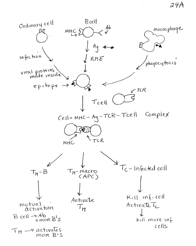
IV. Antigen Presentation & Major Histocompatibility Complex (MHC). See handout 24A. (For better pictures see Purves 19.18).
A. What is MHC?
1. MHC = very variable surface protein. There are 2 main types, and many versions of each type. Each individual has several different genes for each of the two main types of MHC. Each of these genes has 20-40 or even more variants (alleles). Since there are several genes per person and many different alleles of each gene in the population, there is a lot of variation in the actual MHC proteins (and DNA) from person to person. These genes, unlike genes for antibodies and TCR's, do not rearrange during development. So there is variation from person to person, but all cells in a single person have the same MHC genes.
2. Two types of MHC
a. All nucleated cells have MHC I on their surface.
b. Cells of immune system (all APC's) have MHC II on their surface. (Not all T cells have MHC II at all times, & we will assume T cells do not have MHC II.)
B. What are Antigen Presenting Cells (APC's)? APC's = Cells that have antigens bound to MHC on their plasma membranes. How they get their antigens/epitopes and attach them to MHC is shown on the top of 24A. T cells bind to the MHC-Antigen complex, as shown in the middle of the handout. (See Purves 19.17)
1. APC's do not present whole antigens -- APC's present fragments of antigens called epitopes or antigenic determinants. See top 1/2 of 24A & See Purves 19.16
(1). Ordinary cells (not from immune system) present fragments of whatever proteins they are making (+ MHC I). These epitopes come from proteins made inside the APC itself and then partially digested in proteosomes.
(2). Immune system cells (B cells, dendritic cells & macrophages = "classic" APC's) present fragments of whatever they have engulfed or endocytosed (+ MHC II) -- Purves 19.16. These epitopes come from proteins that were originally outside the APC and were partially digested in lysosomes/endosomes.
2. Each APC presents many different epitopes at once (even if they are all derived from a single antigen).
3. How do the epitopes reach the cell surface?
a. The endogenous fragments digested in proteosomes enter the ER (by a special transporter), and combine with newly made MHC molecules (in the ER membrane). The complex is transported to the cell surface through the ER, Golgi, etc. in the same way as any cell surface protein.
b. The exogenous fragments digested in lysosomes/endosomes combine with newly made MHC in the lysosomes/endosomes and the complex reaches the cell surface the same way that used receptors recycle to the surface.
C. Why do you need MHC & APC's?
1. T cells are "MHC restricted;" B cells are not
a. B cells recognize plain Ag = Antibodies bind to Ag in plasma or on bacterial/viral surfaces.
b. T cells recognize only Ag that is bound to MHC on (euk.) cell surface ( Purves 19.17 and handout 24B.)
(1). T cell receptors bind to variable part of MHC-Ag complex = bind to Ag itself
(2). CD4 or CD8 binds to constant part of corresponding MHC.
2. Two types of T's recognize (bind to) Ag associated with different MHC's -- this is how T cells tell immune cells and infected (ordinary) cells apart. See handout 24B.
a. Cytotoxic T's (CD8+) recognize Ag + MHC I (said to be "MHC I restricted") -- note target must have MHC I and Ag.
b. Helper T's (CD4+) recognize Ag + MHC II (said to be "MHC II restricted") -- note target must have MHC II and Ag.
The point: T cells recognize their targets (in part) by the type of MHC they have -- infected cells have MHC I and immune cells have MHC II.
V. Putting it all together -- Purves 19.18 or handout 24A
A. T cell is activated (Middle of 24A)
1. Need binding to APC -- either
a. Binding to classic APC (B cell or phagocytic cell -- macrophage or dendritic cell) to activate TH
(1). In primary response, APC probably a phagocytic cell (not specific for any particular antigen)
(2). In secondary Response, APC likely to be a B cell (with antibody specific for that antigen)
b. Binding to infected cell to activate TC.
2. T cell - APC cell binding requires match
a. APC must have Ag (epitope) + MHC
b. T cell must have TCR that matches Ag, and CD4 or CD8 to match proper MHC.
Note: Picture on handout shows epitope in middle, in between both MHC of APC and TCR of T cell. The epitope is firmly bound to the MHC and stays with the APC when the T cell finishes activation and detaches. The activated T cell now has an empty TCR and will bind to another (B) cell with the same epitope.
3. Cytokines must be provided for activation -- to bind to receptors on T cell.
a. Cytokine (IL-1) from APC needed to activate TH .
b. Different cytokine (IL-2) from TH needed to activate TC. ( This is why you need TH's for cytotoxic T response.)
4. Activation --> clonal expansion (more TH cells) AND more specialization of T cells. These activated T cells can disassociate from the APC and find another cell to "help."
B. What activated TH cell does (see bottom of handout 24A)
1. Humoral Response: Activated TH cell then divides and/or activates a B cell -- activates the same APC that just activated it or finds a new B cell.
2. Cell Mediated Response: Activated TH cell divides and/or helps activate a TC cell (by providing cytokines) -- details of this not discussed.
C. What Activated TC cell does (see bottom of handout): Divides and/or kills an infected cell.
Try problems 13-9 & 13-10. To review all the terminology so far, try 13-11.
VI. How do T and B cells get activated? Wrap Up. See handout 24A. and topic V above. -- This will be discussed next time.
|
What's Activated?** |
Antigen Presenting/Target Cell |
What holds Epitope |
Source of Antigen |
Result |
|
Cytotoxic T |
Infected Cell |
MHCI |
Made in infect. cell |
Killing of Target Cell; |
|
Helper T |
Classic APC (B, macrophage, etc.). |
MHCII |
From outside APC |
Humoral Response as in (1) or (2) below; Mitosis of TH cell to give clone |
|
|
(1) macrophage |
MHCII |
From outside by Phagocytosis |
Activated TH can go on to activate a B; leads to Ab production by B cell |
|
|
(2) B |
MHCII |
From outside by RME |
Mutual Activation of TH and B; Ab production by B cell |
**Note: Activation of lymphocytes also requires appropriate Interleukins. TH cells need IL-1 from the APC's; TC cells need IL-2 from TH and B cells need various IL's; class of Ab made by B cell depends on type of IL it gets.