C2006/F2402 '04 OUTLINE FOR LECTURE #5
Last updated 02/03/04 01:58 PM(c) 2004 Dr. Deborah Mowshowitz , Columbia University, New York, NY
Handouts: Transport of glucose through body -- 5A.; 5B --Receptor Mediated Endocytosis
Here's a nice picture of a channel and an explanation of how K+ channels can be selective.
I. Types of Transport across Membranes, cont.
D. Active Transport (Cases 4 & 5)
1. Curve #1: when it plateaus, [X]in greater than [X]out -- because movement of substance linked to some other energy releasing reaction. (This assumes we are following the reaction Xout --> X in)
2. Curve #2 saturates. Enzyme-like protein involved -- acts as transporter or pump.
3. Energy: Not readily reversible; Keq not = 1 and standard delta G not = zero. Overall reaction usually has large, negative standard delta G because in overall reaction transport of X (uphill, against the gradient) is coupled to a very downhill reaction, as shown in II. B. above. The downhill reaction is either
a. Splitting of ATP (in primary active transport), or
b. Running of some ion (say Y) down its gradient (in secondary active transport).
4. Secondary Active Transport -- How does ATP fit in? Process occurs in 2 steps:
a. Step 1. Preparatory stage: Splitting of ATP sets up a gradient of some ion (say Y), usually a cation.
b. Step 2. Secondary Active Transport Proper: Y runs down its gradient, and the energy obtained is used to drive X up its gradient. See Becker fig. 8-10.
c. Overall: Step (1) is primary active transport; step (2) is secondary and can go on (in the absence of ATP) until the Y gradient is dissipated. Note that step (1) cannot occur at all without ATP but step (2) can continue without any ATP (for a while).
5. How do you tell the two types apart? Primary is directly dependent on splitting of ATP; secondary will continue even in the absence of ATP until the gradient of Y runs down.
Try problem 2-2.
6. Some Examples & Possible mechanisms (see handout 4B, texts & animations for models). Click on links for animations.
|
Example |
Type of Active Transport |
Type of "Port" |
Pictures in Becker |
Pictures in Purves |
|
|
a. |
Primary |
Antiport |
figs. 8-11 & 8-12 (8-10 & 8-11) |
5.12 |
|
|
b. |
Na+/Glucose co-transport |
Secondary |
Symport |
fig. 8-13 (8-12) |
5.13 |
7. Are pumps reversible?
a. Theoretically, all pumps (like Na+/K+ pump) are reversible -- a pump can break down ATP and use the energy to drive ions up their gradient, or (if ion gradient is large enough) ions running down their gradient can provide enough delta G to drive phosphorylation of ADP to ATP. Therefore, proteins that catalyze active transport are sometimes called "ATPases" or pumps, whether their normal function is to hydrolyze ATP or to synthesize ATP.
b. Practically speaking, inside cells, most pumps are irreversible. Most (but not all) individual "pump" proteins work only one way in cells, because the standard delta G for the "usual" direction is very negative. Therefore it takes very high concentrations of products (very high ATP or very high ion concentrations, depending on the reaction) to push the reaction in the "reverse" direction. The concentrations needed to reverse the reaction are not reached in cells, but can be achieved in test tubes (by adding ATP, setting up ion gradients, etc.). So in vitro (in test tubes), but not in vivo (in living cells), you can make the pumps run in either direction. Two examples of important pumps that are reversible (in vitro), but usually run in one direction (in vivo):
(1). In the inner membranes of mitochondria and chloroplasts, chemical or light energy is used via electron transport to set up a proton gradient, which then runs down; driving phosphorylation of ATP. So these systems almost always act to make ATP while ions run down their gradient.
(2). The Na+/K+ pump in the plasma membrane almost always uses up ATP -- this system drives ions up their gradients at the expense of ATP.
For more examples, see Becker table 8-3.
Now try problems 2-3 & 2-5.
II. Putting all the Methods of Transport of Small Molecules Together or What Good is All This?
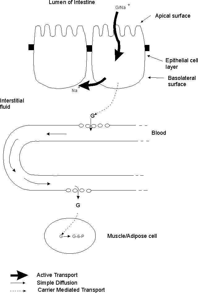
A. How glucose gets from lumen of intestine --> muscle and adipose cells. An example of how the various types of transport are used. (Handout 5A and Becker fig. 11-22) Steps in the process:
1. How glucose exits lumen. Glucose crosses apical surface of epithelial cells primarily by Na+/Glucose co-transport. (2o act. transport)
2. Role of Na+/K+ pump. Pump in basolateral (BL) surface keeps Na+ in cell low, so Na+ gradient favors entry of Na+. (1o act. transport)
3. How glucose exits epithelial cells. Glucose (except that used for metabolism of epithelial cell) exits BL surface of cell (and enters interstitial fluid) by facilitated diffusion = carrier mediated transport. (Interst. fluid = fluid in between body cells.)
4. How glucose enters and leaves capillaries -- by simple diffusion through spaces between the cells. Note: this is NOT by diffusion across a membrane.
5. How glucose enters body cells -- by facilitated diffusion (= carrier mediated transport). Carrier is only "mobilized" that is, inserted into membrane (by fusion of vesicles as explained previously) in some cell types (adipose & muscle) in presence of insulin. Carrier is permanently in cell membrane in other cell types (brain, liver). See below on GLUT transporters.
6. Role of glucose phosphorylation. Conversion of G --> G-6-phosphate traps G inside cells.
For additional examples of the uses of the various types of transport processes, see Becker fig. 8-1 & 8-2.
B. How Glucose Reaches Body Cells -- Another look at handout 5-A. -- The steps in the process are described above in the order in which they occur. Here, the focus is on the various types of transport involved.
1. Role of Active transport -- Needed to get glucose from lumen to inside of epithelial cell.
a. Primary active transport -- Na+/K+ pump keeps intracellular [Na+] low.
b. Secondary active transport -- Glucose enters epithelial cells by Na+/Glucose co-transport
2. Role of Passive Transport & Phosphorylation
a. Passive Transport -- Used to move glucose the rest of the way -- out of epithelial cells, in & out of capillaries, and into body cells.
b. Phosphorylation of glucose -- Used in the body cells to keep the free glucose level at the "end of the road" low, and ensure that the glucose gradient is "downhill" from epithelial cells to capillaries to body cells.
3. Role of Diffusion: Glucose and other small molecules (but not macromolecules) diffuse in and out of capillaries through the liquid filled spaces between the cells, not by diffusing across the cell membrane. Note that proteins are too big to enter or leave capillaries this way.
4. Role of GLUT transporters (another protein/gene family)
a. GLUT proteins are responsible for carrier mediated transport of glucose. All passive glucose transport across membranes depends on a family of proteins called GLUT 1, GLUT 2, etc. This family of genes and transport proteins is responsible for all carrier mediated transport of glucose.
b. Different family members (genes and proteins) are expressed in different cell types. GLUT 1 protein is found in plasma membrane of RBC & most other cells, GLUT 2 protein on BL surface of intestinal epithelial cells, GLUT 4 protein in muscle and adipose, etc. (Note all genes for all proteins are present in all these cell types -- DNA is the same!)
c. All the genes and corresponding proteins are similar, but have significant structural and functional differences. This is another example of a gene/protein family. All the proteins have a similar overall structure -- 12 transmembrane segments, COOH and amino ends on intracellular side of membrane, etc.
d. Position & Action of GLUT 4 is insulin dependent. GLUT 4 is the only insulin dependent member of the family. Insulin triggers insertion of GLUT 4 protein into the plasma membrane, by triggering vesicle fusion, as explained last time. All the other proteins are located constitutively in their respective membranes.
e. Direction of transport. Note that one member of this family (GLUT 2) is responsible for ferrying glucose OUT of epithelial cells; different members are responsible for helping glucose ENTER most other cells. All family members bind glucose on one side of the membrane, change conformation and release glucose on the other side of the membrane. Which way the glucose goes (overall) depends on the relative concentrations of glucose on the two sides of the respective membrane, not on which GLUT protein is used.
Try problem 2-9.
III. Ways that Big Molecules Enter Cells.
A. Pinocytosis = bulk phase endocytosis; no receptor. Take in random samples of surrounding fluid. See Becker fig. 12-13.
B. Phagocytosis -- in specialized cells only -- extensions of cells (pseudopods) reach out and engulf solids. See Becker fig. 12-14. Vesicle that is formed is called a phagocytic vesicle (or vacuole) or phagosome.
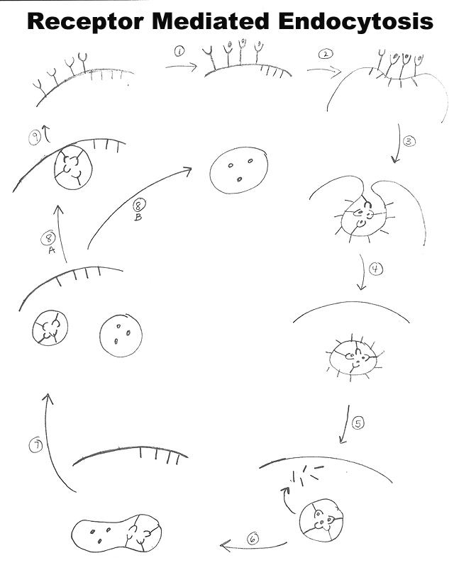
C. RME = receptor mediated endocytosis. Cells take in specific substances from surrounding fluid using a receptor. See Becker fig. 12-15 (diagram) & 12-16 (micrograph) or Purves 5.15 (5.15 & 5.16). Different cell types have different combinations of receptors.
IV. RME -- Receptor Mediated Endocytosis
A. General and/or important Features.
1. Receptors -- Need specific receptor for each substance (or class of closely related substances) to be transported 2. Concentrates substances transported -- moves them up their gradient. 3. Requires energy (not clear exactly which stages in process use ATP or GTP.) Energy must be required because substances move against their gradients.
4. Role of clathrin -- A protein is needed to deform membrane and allow vesicles to form -- provides a coat. (See Becker figs. 12-15 to 2-18 and/or Purves 5.15.)
Other proteins are required as well, but will not be discussed.a. Clathrin is coat protein for vesicles forming from plasma membrane and trans-Golgi. (trans side of Golgi = "far end" = side away from nucleus and ER = last part that proteins travel through as they are processed in Golgi. Also called "TGN" for "trans Golgi Network." See Purves 5.15 or Becker fig. 12-8 for labeled pictures.)
b. Budding of other membranes involve different "coat" proteins. Best known are COPI & COPII which are involved in ER-Golgi transport. (See Becker for details if interested. Types of coats are summarized in table 12-2.)
5. It's a cycle -- Exocytosis balances endocytosis so cell surface area stays the same. See Purves 5.18 or Becker fig. 12-15. For LDL receptor, it takes about 10-20' for one "round trip."
6. Topology -- material can enter and/or exit cell without being in contact with cytoplasm. Material can remain inside a vesicle or outside cell at all times.
B. Stages of Cycle (Numbers match steps on handout 5A.) Click here for animation.
(1). Receptors bind material
(ligand) to be internalized(2). Receptors are in (or migrate to) coated pits (clathrin
-coated parts of membrane)(3). Membrane starts to invaginate to form coated vesicle
. A single vesicle can contain more than one type of receptor plus ligand.(4). Coated vesicle forms
(pinching off of vesicle may be an energy requiring step)(5). Uncoating occurs relatively quickly
(uncoating may require energy)(6). Vesicle is acidified, and sorting of receptor(s) and ligand(s) begins.
- A single endosome may contain many different receptors and ligands, and different ones are sorted differently. (Some examples are given in detail below.)
- The uncoated, acidified vesicle can be called an endosome, early endosome, or a sorting vesicle. (Terminology varies -- usage of terms early endosome, late endosome etc. differs in dif. texts.)
- Acidification requires energy to run proton pump -- to move H+ into vesicle at expense of ATP.
Note: Details of sorting and recycling -- the remaining steps -- vary with material endocytosed. More details are below for individual cases.
(7). Endosome splits
. The substance we are following, and/or its receptor, can end up in either half. (In example shown on handout, one half gets the receptor and one half gets the ligand, as is the case for LDL.)Note: endosome may not simply split in one step; process of sorting may be gradual. Pieces of different composition may gradually bud off as internal composition of remainder changes.
(8). What Happens to the Different Parts of the Endosome?
8A. Fate of vesicle with materials to be recycled (receptors and/or carriers) -- this vesicle fuses with plasma membrane. (In case of LDL, this vesicle would contain the receptor for LDL.)
8B. Fate of vesicle with material that was endocytosed -- Vesicle delivers contents to appropriate cell compartment. (For LDL fuses with lysosomes so material is degraded.)
(9). Exocytosis occurs
-- returns receptors and/or other components to the plasma membrane or outside of cell.
Try Problem 2-6.
1. LDL (Low density lipoprotein) -- more details. See Becker, Box 12B or Purves 5.16 (5th ed.)C. Some Specific Examples
a. What is it? A particle containing cholesterol esters + some other lipids + a protein (carrier). Particle is covered by monolayer of phospholipid plus some unesterified cholesterol.
2. Fe/Transferrinb. Why LDL? Cholesterol is insoluble in blood. (Too hydrophobic.) Need a way to ferry cholesterol through blood and into cell -- Cholesterol transport requires formation of particle with hydrophilic surface; binding to cell surface receptor requires a protein to bind to receptor.
(1). Ligand = what actually binds receptor = protein part of LDL = carrier protein
(2). What cell actually needs is the cholesterol part.
c. Receptor, but not carrier, is recycled. Note: there are 2 separate proteins here that are easily confused
(1) Receptor protein on the cell surface = LDL receptor = binds LDL and allows uptake of cholesterol
(2) Carrier protein = ligand for LDL receptor = part of LDL and helps carry cholesterol through the blood.
d. Receptor and carrier are separated inside sorting vesicles/endosomes
e. Need lysosomes to degrade carrier and release cholesterol (cholesterol esters in LDL must be split for cholesterol to be used).
(1). How LDL reaches lysosomes: vesicles/endosomes holding substrate fuse with pre-existing lysosomes and/or endosomes fuse with vesicles from Golgi carrying newly made hydrolases to form new lysosomes. (More details on how hydrolases pass through the Golgi and are targeted to lysosomes to be discussed later.)
(2) Current terminology: relationship of early endosomes, late endosomes & lysosomes. Note: Most of this is FYI. In this course, the term "endosomes" will be used for both early and late endosomes.
(a). Early endosome = An acidified vesicle that is early in the pathway that leads to lysosomes. Includes both materials to be sent to lysosomes (hydrolases or substrates) and materials to be recycled for reuse (receptors, etc.). Early endosomes can include material from either or both of the following:
(i) Vesicles from invagination of plasma membrane carrying newly endocytosed material, and
(ii). Vesicles coming from Golgi carrying newly made hydrolases (more on this later).
(b). Late endosome = vesicle containing hydrolytic enzymes (not yet activated) plus potential substrate. More acidic than early endosome. Material not destined for lysosomes has been jettisoned.
(c). Lysosomes = vesicle containing active hydrolytic enzymes and substrate. More acidic than late endosome. Formed by maturation of late endosome and/or fusion with pre-existing lysosome.
(3). Older terminology found in some texts (FYI only):
(a). Primary lysosome = vesicle with enzymes only.
(b). Secondary lysosome = enzymes + substrate = result of fusion of primary lyso. + another vesicle containing substrate.
f. Function of uptake -- to supply a nutrient (cholesterol).
a. What is transferrin? Fe needs carrier protein (like cholesterol does) for transport and binding to receptor; carrier (= ligand for cell receptor) is called transferrin b. Both carrier & receptor are recycled c. No lysosomes needed -- iron diffuses out of endosome; no protein is degraded d. Carrier and receptor separate outside cell after recycled(1). Fe/transferrin binds to receptor at neutral pH and enters cell by RME. (2). Inside cell, Fe diffuses out of vesicle into cytoplasm, leaving apo-transferrin stuck to receptor ("apo" means without ligand or cofactor).(3). Apo-transferrin (without Fe) sticks to receptor at low pH (in endosome) but separates at neutral pH (outside cell). This is contrary to usual behavior -- Most ligands stick to receptors at neutral pH but separate at low pH found in endosome.
(4). Note that apo-transferrin and Fe/transferrin have different affinities for the receptor at neutral pH. Under these conditions, Fe/transferrin binds to the receptor, and apo-transferrin separates from the receptor.
e. Function of uptake -- to supply a nutrient (Fe).
3. EGF (Epidermal Growth Factor)a. No carrier required; EGF (a protein -- unlike Fe or cholesterol) alone binds to receptor; EGF = signaling molecule = ligand for cell surface receptor & substance that will be transported into the cell.
b. Function of uptake -- to regulate signaling -- turn off signal and down regulate receptors (reduce # of cell surface receptors).
c. Receptor not recycled -- Ligand (signal molecule) and receptor degraded together.
d. Need lysosomes (to degrade both receptor and ligand)
D. For Reference: Compare & Contrast for the examples described above for transport of X
|
Transferrin |
LDL |
EGF |
|
|
Lysosomes Involved? |
No |
Yes |
Yes |
|
Carrier/Ligand Fate |
Recycled |
Digested |
No carrier |
|
Receptor Fate |
Recycled |
Recycled |
Probably Digested |
|
What's carried in (what is X)? |
Fe |
Cholesterol |
Growth Factor |
|
Function of X |
Metabolism (Fe is cofactor for many proteins) |
Metabolism (cholesterol is a component of cell membranes; used for hormone synthesis) |
Signal |
|
Ligand (What binds receptor?) |
Transferrin |
LDL (protein part) |
EGF |
|
Do ligand & receptor separate inside cell? |
No |
Yes |
No |
|
Where do ligand & receptor separate? |
Outside the cell |
In endosomes |
Not separated -- both degraded |
Next Time: How do we know where proteins go? How are proteins targeted to
to their proper destination within the cell?