C2006/F2402 '05 Outline for Lecture 20 -- (c) 2005D. Mowshowitz -- Lecture updated 04/12/05. Minor changes made after the morning lecture.
Handouts: 20A Circuits, 20B CNS/PNS, 20C PNS (Extra copies in boxes outside Dr. M's office; 7th floor Mudd.)
This lecture will fill in some features of the nervous system not covered by Dr. Firestein. Muscle will be covered next time. Parts of topics I & II have already been covered by Dr. F, but are included here for reference. Note that the handout on the AP incorrectly labels the refractory periods. The absolute refractory period coincides more or less with the spike; the relative refractory period follows after the spike.
1. Sensors Purves has a whole chapter on Sensory Systems. (Chapter # depends on which edition of the text you have.) Dr. F went over the overall principles that are covered in the first few pp of the chapter. Today we will do a few details of specific detection systems. We will not cover all the details of each system.
A. Two types of Receptor Cells = sensory cells = special cells with molecular receptors for detecting stimuli
1. Modified neuron capable of generating AP itself.
2. Cell that cannot generate an AP itself, but releases transmitter and triggers AP in next cell. This type of receptor cell can be a modified neuron or epithelial cell.
Question to ask yourself: What type of channels does a cell need in order to generate an AP?
B. Receptor Proteins.
1. Special cells contain receptor proteins for stimuli (pressure, light, heat, chemicals, etc.).
2. How do protein receptors detect stimuli?
a. Stimuli --> Change in conformation of receptor --> open or close channels in membrane --> change in polarization of membrane.
b. How are channels opened or closed? See Purves 45.1
(1) Directly -- receptor is part of a channel. Examples: receptors for mechanical stimuli (touch, hearing, balance), & temperature (heat/cold). Sensor uses an ionotropic receptor. (See section below on receptors of PNS.)
(2) Indirectly -- through 2nd messengers. Examples: receptors for chemicals (taste, smell, etc.), electromagnetic radiation (vision). Sensor uses a metabotropic receptor.
C. Receptor Potentials are Graded
1. Response to stimulus is graded. Stimulus --> local graded response. The more stimulus, the more channels open (or close), and the bigger the graded potential (bigger depolarization or bigger hyperpolarization).
2. Terminology -- graded response in receptor cell is called a generator potential or receptor potential.
D. How Does Graded response generate an AP? -- see Purves 45.2.(6th ed. only) for the two methods.
1. In modified neuron. Graded (generator*) potential triggers AP in same cell (if stimulus is over threshold) --> input to CNS.
a. Example #1 -- direct (ionotropic receptor) -- stretch.
Stimulus = stretch on nerve endings --> open channels --> depolarize to threshhold --> AP.b. Example #2 -- indirect (metabotropic receptor) -- smell (olfaction).
Stimulus = Ligand (odorant) --> Receptor --> G protein --> adenyl cyclase --> cAMP up --> open cation channel (cyclic nucleotide gated channel) --> depolarize cell --> AP.
Question: Where will the action potential start? In what part of the cell? See Purves 45.2 (45.3)
2. In separate receptor cell: Graded (receptor*) potential triggers release/inhibition of transmitter by receptor cell -- amount of transmitter released is proportional to stimulus. Transmitter generates IPSP or EPSP in neuron (next cell = post synaptic cell). Transmitter triggers AP in post synaptic neuron if stimulus is over threshold --> input to CNS. Example: vision, taste, balance, hearing.
* In older editions of Purves, the terms "generator potential" and "receptor potential" are used to refer to these two different cases respectively. Most texts stick to "receptor potential" or use the two terms interchangeably.
Which type of receptor cell are you dealing with in problem 8-16?
3. Details for Vision -- Example #3 -- see Purves 45.12 to 45.14 (45.14 to 45.17) for signal transduction events. (This is really a 3 cell system, not a 2 cell system. 3rd cell, not 2nd, generates an AP.)*
a. Receptor cell (in the dark) releases an inhibitory transmitter, so post synaptic neuron remains hyperpolarized and does not fire.
In dark: receptor cell --> cGMP --> open cation channel --> cell depolarized --> releases inhibitory NT --> IPSP --> no AP in subsequent cells --> no message to brain.
b. Binding of photons to pigment in receptor cell blocks release of inhibitory neurotransmitter. Post synaptic neuron depolarizes and fires an AP.
In light: Receptor (rhodopsin w/ cis retinal) binds photon --> rhodopsin w/ all trans retinal --> G protein --> activates phosphodiesterase (PDE) --> cGMP degraded --> cation channel closes, cell hyperpolarizes --> less (inhibitory) transmitter release --> AP in subsequent (3rd) cell --> message to brain. For more details see Purves Ch. 45.*Receptor cell releases NT --> bipolar cells, which release NT --> ganglion cells. (Bipolar cells cannot fire an AP, but can excite or inhibit ganglion cells.) Ganglion cells, which have input from multiple bipolar cells, can fire an AP.
E. All stimuli (whatever the modality) give same message to CNS (= AP's). If AP is all or nothing, how do you know which stimulus it was? And how much? See notes for lecture 19.
1. Number, frequency of AP's indicate length (duration) and strength (intensity) of stimulus.
2. Wiring (what part of brain is stimulated) = labeled lines = indicates location of stimulus and type (modality) of stimulus -- taste, stretch, etc. (Take a very sharp pencil and tap your upper lip. What sensors did you trip off?)
To review sensors, try Problem 8-16 . To review electrical communication overall, try 8-15.
II. Nerve-Nerve Synapses, cont. For nice overall pictures see Purves 44.13 (44.16) or Becker fig. 9-21 & 9-23. Some of this is review, but is included for clarity. What determines if a nerve impulse will be passed on to the next neuron?
A. Presynaptic Side -- Transmitters -- See Purves Table 44.1 & Becker fig. 9-22
1. One major type of transmitter per synapse (released from pre-synaptic side)
a. In CNS, many diff. transmitters. Usually amino acids or their derivatives. Major ones are glutamate (excitatory) & GABA (inhibitory).
b. In PNS usually norepinephrine (NE) or acetyl choline (AcCh).
2. One transmitter per Neuron. Usually only one transmitter released by any neuron. Therefore one major transmitter released -- the same one -- at all synapses made by that neuron.
3. Effects of transmitter can vary. Some transmitters always excit. or inhib; other transmitters vary (depends on if have one or more types of receptor for that transmitter )
4. Getting rid of transmitters
a. Transmitter doesn't remain in cleft for long.
b. Different methods for getting rid of dif. transmitters
(1). Diffusion away from synapse
(2). Destruction by enzymes in synapse. For ex, acetyl choline esterase (AcChE) in cleft breaks up AcCh (Choline is reused). See Purves fig.44.14 (44.15)
(3). Reuptake by endocytosis -- NE, serotonin removed by reuptake
c. Many drugs affect release/fate of transmitters; for ex.
(1). Prozac prevents serotonin reuptake -- transmitter stays around longer --> more stimulation.
(2). Malathion (insecticide) & nerve gas block AcChEsterase --> continuous stimulation --> spasms
B. Post Synaptic Side --PSP's = post synaptic potentials = small change in potential due to release of transmitter at a single synapse.
1. Can be inhibitory (generate an IPSP) or excitatory (generate an EPSP)
a. Inhibitory -- causes hyperpolarization or stabilizes existing negative polarization due to opening of K+ or Cl- channels. Either K+ goes out or Cl- comes in.
b. Excitatory -- causes depolarization due to opening of cation channels; Na+ in>> K+ out.
2. Any given synapse is excitatory or inhibitory -- What determines it?
One type of receptor for neurotransmitter at each synapse -- Receptor determines which kind of synapse it is -- excitatory or inhibitory. Same neurotransmitter can be excitatory or inhibitor at different synapses.)
Overall: One receptor/neurotransmitter pair per synapse. (Receptors of PNS are in table below.)
3. Features of PSP's (To compare to AP's)
a. PSP's are graded (size is proportional to stimulus as with receptor potentials), not all or none. (Unlike action potentials.)
b. PSP's are local -- die out if don't reach threshold. (Not regenerated like AP's.)
c. PSP's are caused by opening/closing of ligand gated channels. (What kind of channel is needed for AP's?)
To review IPSP's and EPSP's try problem 8-10.
C. Post Synaptic Side -- Summation -- See Purves fig. 44.15 (44.17) or Becker 9-26 & 9-27.
1. Inputs (IPSP's & EPSP's) to cell body/dendrites are summed -- changes spread around cell body to axon hillock (or die out)
2. No AP in cell body. No voltage gated channels in cell body so no AP generated there
3. Axon Hillock. Voltage gated channels begin at axon hillock (some texts call this the "trigger zone") so AP starts there. See Becker fig. 9-16 or Purves 44.15 ( 44.17).
4. Inputs summed over space and/or time -- need to depolarize past threshold at axon hillock to --> AP
a. Spatial summation: Multiple EPSP's delivered at different spots can add up --> AP
b. Temporal summation: Multiple EPSP's delivered close enough together in time can add up --> AP
c. Why you need summation:
(1). A single EPSP is not enough to --> AP
(2). IPSP's & EPSP's are summed: net effect depends on both inhibitory and excitatory input. (See circuits below for why you need this.)
Review Problem 8-8, parts A to H, and recitation problem 11-1.
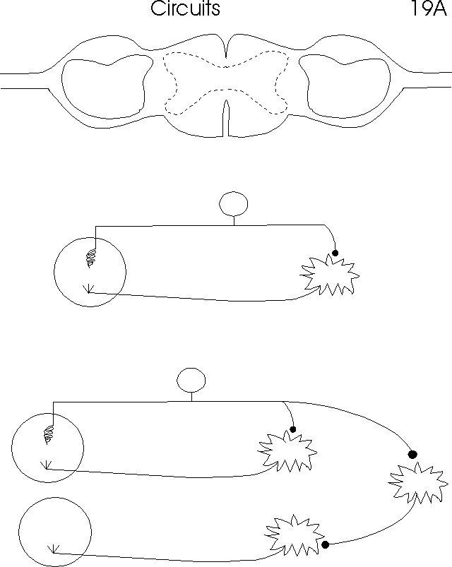
III. Circuits -- how nervous system is organized
A. Simple circuits -- see handout 20A or Purves 46.3 (46.4)
1. One synapse, 2 neurons -- monosynaptic circuit -- how sensory neuron signals an effector.
2. Circuit with multiple synapses -- how antagonistic muscles are controlled. (Signal to skeletal muscle is always +; + = signal to contract; no signal = relax.)
3. Role of brain -- adds up/down (as vs. in/out) component
4. FYI: Where is all this located? see Purves 46.3 (46.4) -- top of handout 20A; not discussed in class.
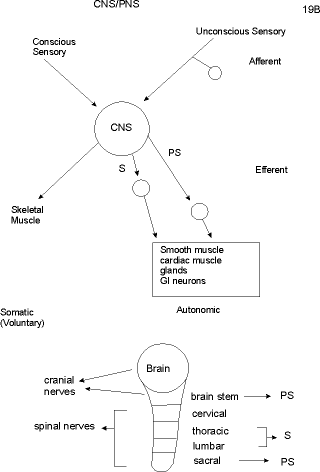
B. How is NS organized overall? See handout 20 B or Becker fig. 9-1 or Purves 46.1 (46.2)
1. CNS
a. CNS = brain + spinal cord
b. Interneurons. Most neurons of CNS are interneurons (99%)
c. White matter = axons
d. Grey matter = cell bodies, interneurons, and dendrites
2. PNS -- Names of Divisons
a. Afferent vs Efferent.
(1) Afferent =carrying info into the CNS
(2) Efferent = carrying info away from the CNSb. Efferent subdivided into: Somatic vs autonomic
(1) Somatic = controls skeletal muscle
(2). Autonomic = controls everything elsec. Autonomic subdivided into: Parasympathetic (PS) vs Sympathetic (S)
Try problem 8-8, part I.
C. How do PS and S co-operate? (See Purves 46.10 (46.11)) What do they do?
1. What do they innervate?
a. Many organs innervated by both
b. Some organs innervated (stimulated) by only one
(1). liver, sweat glands -- S only
(2). tears -- PS only
2. What results does stimulation produce?
a. Not always S excites; PS inhibits. Ex: salivation -- S inhibits; PS excites
b. Usually: S --> response needed in a crisis; PS --> response needed in relaxed state.
c. Examples:
(1). S --> heart rate up; liver releases glucose; bladder relaxes (to hold more);
(2). PS --> heart rate down, digestion, salivation up.
d. See Stress Response described previously. Note all nerves involved in stress response (and adrenal medulla) are part of sympathetic division.
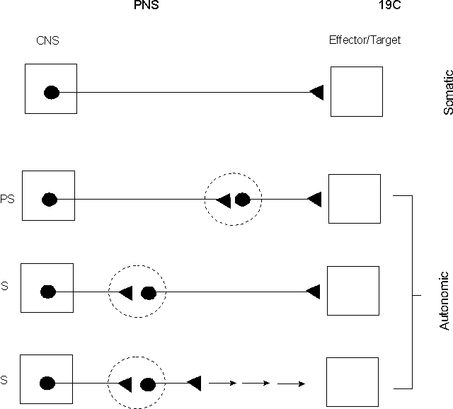
D. General Set up of wiring of efferent PNS -- see handout 20C
1. First neuron -- same in Somatic and autonomic.
a. body in CNS
b. releases AcCh (nicotinic receptor on effector/next neuron)
2. Second neuron (post ganglionic) -- found in autonomic only
a. Body in ganglion
b. Releases AcCh (muscarinic receptor on effector) or NE
c. Adrenal medulla = second neuron with a short axon
Try problem 8-8 part J.
E. Receptors in the PNS
1. Response to particular transmitter depends on receptor as with hormones.
2. Receptors can be direct or indirect (as with hormones or sensory receptors)
a. Direct or ionotropic: Receptors (for transmitters) are ligand-gated ion channels; relatively fast. For examples see Purves 44.17 (44.19) or Becker 9-25. Also see many pictures in both books of neuromuscular junction.
b. Indirect or metabotropic: Receptors are coupled to G proteins, generate 2nd messengers ---> open or close ion channels or have other effects (i.e. act like hormones); relatively slow. See Purves 44.16 (44.18)
3. Agonists and antagonists used as common tools to study receptors, as with hormones. Some receptors named by their most common agonist or antagonist -- see table below.
4. Examples -- Major types of receptors in the PNS
|
Basic Type of Receptor |
Transmitter |
Detailed Type of Receptor |
Mechanism |
Agonist |
Antagonist |
Typical effect |
Cholinergic |
Acetyl Choline |
Nicotinic |
Direct (It's a Na+/K+ channel) |
Nicotine |
Curare |
Contract skeletal muscle |
Cholinergic |
AcCh |
Muscarinic |
Indirect; 2nd messenger & effect varies |
Toxin from Amanita muscara (muscarine) |
Atropine |
Slow heart beat |
Adrenergic |
Epinephrine (adrenalin) & NE; NE> E |
Alpha* |
Indirect; 2nd messenger & effect varies |
|
|
Contract smooth muscle |
Adrenergic |
Epi. & NE; E> NE |
Beta* |
Indirect; cAMP is 2nd messenger |
Asthma drugs |
Beta blocker |
Relax smooth muscle (bronchodilates); increase heart beat |
See Becker fig. 23-18 for diagram of action of adrenergic receptors
*Both alpha and beta have subtypes that differ in location, mechanism & effect; same receptors used for epinephrine acting as a hormone. (See also handout 13A.)
** This is the receptor at the neuromuscular junction. This is the AcCh receptor Becker (in Ch. 9, fig. 9-25) and many others mean when they talk about "THE" acetyl choline receptor.
Look at problem 8-16, part C. Assume you are looking at a standard synapse between two nerves that uses AcCh as a transmitter. What are the answers to parts 1-5? What effects will you expect on transmission at a standard synapse? (Note -- this problem as written does not refer to a standard synapse between two nerves. However the answers in the back of the book are still okay except for part 4. What is the right answer to part 4 for a standard synapse?)
F. PS vs S -- factors to compare and contrast (see handouts and Purves fig. 46.10 (46.11))
1. Where are ganglia? Near spinal cord or target organs?
2. Where are bodies in CNS = where in spinal cord/brain stem do axons of first neuron come from? See bottom of 20B.
3. What type of synapse with effector? Cholinergic or adrenergic?
4. General effect -- stress or relaxation?
To review the organization of the nervous system, try Problems 8-12 to 8-14.
Next time: How do nerves and muscles work to give contractions?