C2006/F2402 '06 OUTLINE FOR LECTURE #5
Last updated 02/01/06 10:30 AM(c) 2006 Dr. Deborah Mowshowitz , Columbia University, New York, NY
Handouts: 5ATransport of glucose through body -- , 5B -- Models for Active Transport , 5C--Receptor Mediated Endocytosis
I. Transport of Small Molecules, cont.
A. Channels
1. Gating
a. Some Channels are gated = % time any particular gate is open is controlled (but each individual gate is either open all the way or shut).
(1). Ligand gated -- opens or shuts in response to ligands (= chemicals that bind to substance under discussion). Typical substances that open ligand gated channels are hormones, neurotransmitters, etc. For a picture see Purves 5.9.
(2). Voltage gated -- opens or shuts in response to changes in voltage. Allows transmission of electrical signals as in muscle and nerve -- see Becker figs. 13-8 & 13-9 (9-9 & 9-10).
(3). Mechanically gated -- opens or shuts in response to pressure. Important in touch, hearing and balance.
b. Some channels are open all the time (ungated); An example = K+ leak channels. These allow a little K+ to leave or "leak out" of cells, causing cells to have a slight overall negative charge. This is critical to conduction of impulses by nerve and muscle as will be explained in detail later. Why do leak channels only allow "a little" K+ to leave? Why isn't the concentration of K+ on both sides of the membrane the same? See below.
2. Most channels are ion channels -- transport charged particles, not neutral molecules. This raises new energy considerations:
a. Role of charge: If X is charged, need to consider both chemical gradient & voltage (charge gradient). These can both "push" ions the same way or push in opposite directions.
b. Result of charge: Keq not usually equal to 1 here -- Curve #1 plateaus when chemical gradient and voltage are balanced (not necessarily at [X]out = [X]in). Example: K+ ions stop leaking out of the cell and you reach equilibrium for K+ when the charge difference across the cell membrane (which pushes K+ in) balances out the concentration difference across the membrane (which pushes K+ out).
See problem 2-6, A. Can you rule out transport through a channel?
B. Active Transport -- How does it work? See previous lecture for links to animations & references to pictures in texts.
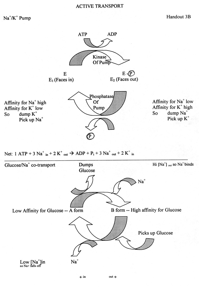
1. An example of primary active transport: Na+/K+ pump (See handout)
a. Pump/enzyme has 2 major forms: one faces in (E1), one faces out (E2).
b. Forms have different affinities for K+ and Na+. (See handout)
c. Role of Phosphorylation: addition/removal of phosphate switches the pump from one form to the other.
d. Role of kinases & phosphatases
(1). This is an example of the use of kinases & phosphatases (for addition/removal of phosphates) to regulate protein activity by reversible covalent modification.
(2). Result of enzyme action in this case:
Kinase activity (catalyzes phosphorylation) flips pump "out" ( E1 --> E2)
Phosphatase activity (catalyzes removal of phosphate) flips pump "in" (E2 --> E1)
e. Location of Enzymes: kinase and phosphatase are part of the pump itself. Not separate proteins.
f. How one cycle goes:
Binding of Na+ on inside activates kinase. Kinase phosphorylates pump. Flips pump out, dumps Na+, picks up K+.
Binding of K+ on outside activates phosphatase. Phosphatase de-phosphorylates pump. Flips pump in, dumps K+, picks up Na+.
g. Stochiometry: 3 Na+ out per 2 K+ in. Some of charge differential balanced by anion transport. Cells are negative on inside relative to outside, but most of charge imbalance is NOT due to pump. (Most charge imbalance is due to K+ leak channels as explained above.)
2. An example of secondary active transport: Na+/Glucose co-transport
a. Enzyme has 2 forms with different affinities for glucose. Either form can face in or out.
b. Role of Na+: Binding of Na+ switches the protein from one form to the other; alters affinity for glucose.
c. Conformational Change: Binding of both glucose & Na+ probably flips protein so it faces the "other" way; loss of both glucose & Na+ does the same -- flips protein so it faces the "other" way.
d. Direction of glucose transport & reversibility: Either form (with or w/o Na+) can face in or out. In normal cell, glucose always goes into the cell with Na+. Why?
3. ABC Transporters
a. Large super family of transporters. Wide range of substances, not just cations, can be transported. Different proteins transport different classes of substances. All ABC transporters are similar in structure.
b. Type of primary active transport. ATP binds to cytoplasmic protein domain called an ATP binding cassette. ATP hydrolysis drives movement of substance being transported.
c. Examples:
(1). MDR protein = multiple drug resistance protein. Pumps many different hydrophobic drugs out of cells; causes lack of response to many cancer treatments.
(2). Protein that controls type of ear wax, and presumably some other (important function), such as the amount of sweat. See article.
d. Important Related Protein: Protein defective in cystic fibrosis belongs to same super family and is similar in structure, but acts as a channel (for Cl-), not a transporter. ATP probably opens and closes channel. See Becker Box 8B.
4. Are pumps reversible?
a. Theoretically, all pumps (like Na+/K+ pump) are reversible -- a pump can break down ATP and use the energy to drive ions up their gradient, or (if ion gradient is large enough) ions running down their gradient can provide enough free energy to drive phosphorylation of ADP to ATP. Therefore, proteins that catalyze active transport are sometimes called "ATPases" or pumps, whether their normal function is to hydrolyze ATP or to synthesize ATP.
b. Practically speaking, inside cells, most pumps are irreversible. Most (but not all) individual "pump" proteins work only one way in cells, because the standard free energy for the "usual" direction is very negative. Therefore it takes very high concentrations of products (very high ATP or very high ion concentrations, depending on the reaction) to push the reaction in the "reverse" direction. The concentrations needed to reverse the reaction are not reached in cells, but can be achieved in test tubes (by adding ATP, setting up ion gradients, etc.). So in vitro (in test tubes), but not in vivo (in living cells), you can make the pumps run in either direction. Two examples of important pumps that are reversible (in vitro), but usually run in one direction (in vivo):
(1). In the inner membranes of mitochondria and chloroplasts, chemical or light energy is used via electron transport to set up a proton gradient, which then runs down; driving phosphorylation of ATP. So these systems almost always act to make ATP while ions run down their gradient. (Diff. proteins used in the two organelles.)
(2). The Na+/K+ pump in the plasma membrane almost always uses up ATP -- this system drives ions up their gradients at the expense of ATP.
For more examples, see Becker table 8-3.
Now try problems 2-3 & 2-5.
II. Putting all the Methods of Transport of Small Molecules Together or What Good is All This?
A. How glucose gets from lumen of intestine --> muscle and adipose cells. An example of how the various types of transport are used. (Handout 5A and Becker fig. 11-22 in 5th ed; not in 6th.) Steps in the process:
1. How glucose exits lumen. Glucose crosses apical surface of epithelial cells primarily by Na+/Glucose co-transport. (2o act. transport)
2. Role of Na+/K+ pump. Pump in basolateral (BL) surface keeps Na+ in cell low, so Na+ gradient favors entry of Na+. (1o act. transport)
3. How glucose exits epithelial cells. Glucose (except that used for metabolism of epithelial cell) exits BL surface of cell (and enters interstitial fluid) by facilitated diffusion = carrier mediated transport. (Interst. fluid = fluid in between body cells.)
4. How glucose enters and leaves capillaries -- by simple diffusion through spaces between the cells. Note: this is NOT by diffusion across a membrane.
5. How glucose enters body cells -- by facilitated diffusion (= carrier mediated transport). Carrier is only "mobilized" that is, inserted into membrane (by fusion of vesicles as explained previously) in some cell types (adipose & muscle) in presence of insulin. Carrier is permanently in cell membrane in other cell types (brain, liver). See below on GLUT transporters.
6. Role of glucose phosphorylation. Conversion of G --> G-6-phosphate traps G inside cells.
For additional examples of the uses of the various types of transport processes, see Becker fig. 8-1 & 8-2.
B. How Glucose Reaches Body Cells -- Another look at handout 5-A. -- The steps in the process are described above in the order in which they occur. Here, the focus is on the various types of transport involved.
1. Role of Active transport -- Needed to get glucose from lumen to inside of epithelial cell.
a. Primary active transport -- Na+/K+ pump keeps intracellular [Na+] low.
b. Secondary active transport -- Glucose enters epithelial cells by Na+/Glucose co-transport
2. Role of Passive Transport & Phosphorylation
a. Passive Transport -- Used to move glucose the rest of the way -- out of epithelial cells, in & out of capillaries, and into body cells.
b. Phosphorylation of glucose -- Used in the body cells to keep the free glucose level at the "end of the road" low, and ensure that the glucose gradient is "downhill" from epithelial cells to capillaries to body cells.
3. Role of Diffusion: Glucose and other small molecules (but not macromolecules) diffuse in and out of capillaries through the liquid filled spaces between the cells, not by diffusing across the cell membrane. Note that proteins are too big to enter or leave capillaries this way.
4. Role of GLUT transporters (another protein/gene family)
a. GLUT proteins are responsible for carrier mediated transport of glucose. All passive glucose transport across membranes depends on a family of proteins called GLUT 1, GLUT 2, etc. This family of genes and transport proteins is responsible for all carrier mediated transport of glucose.
b. Different family members (genes and proteins) are expressed in different cell types. GLUT 1 protein is found in plasma membrane of RBC & most other cells, GLUT 2 protein on BL surface of intestinal epithelial cells, GLUT 4 protein in muscle and adipose, etc. (Note all genes for all proteins are present in all these cell types -- DNA is the same!)
c. All the genes and corresponding proteins are similar, but have significant structural and functional differences. This is another example of a gene/protein family. All the proteins have a similar overall structure -- 12 transmembrane segments, COOH and amino ends on intracellular side of membrane, etc.
d. Position & Action of GLUT 4 is insulin dependent. GLUT 4 is the only insulin dependent member of the family. Insulin triggers insertion of GLUT 4 protein into the plasma membrane, by triggering vesicle fusion, as explained above. All the other proteins are located constitutively in their respective membranes.
e. Direction of transport. Note that one member of this family (GLUT 2) is responsible for ferrying glucose OUT of epithelial cells; different members are responsible for helping glucose ENTER most other cells. All family members bind glucose on one side of the membrane, change conformation and release glucose on the other side of the membrane. Which way the glucose goes (overall) depends on the relative concentrations of glucose on the two sides of the respective membrane, not on which GLUT protein is used.
Try problem 2-9.
III. Ways that Big Molecules Enter Cells -- types of Endocytosis. In all cases net effect is that cell membrane folds in and pinches off, forming a vesicle in the cytoplasm that contains material from the outside.
A. Pinocytosis = bulk phase endocytosis; no receptor. Cells take in random samples of surrounding fluid containing a random selection of extracellular substances. See Becker fig. 12-13.
B. Phagocytosis -- in specialized cells only -- extensions of cells (pseudopods) reach out and engulf solids. See Becker fig. 12-14. Vesicle that is formed is called a phagocytic vesicle (or vacuole) or phagosome.
C. RME = receptor mediated endocytosis. Cells take in specific substances from surrounding fluid using a receptor. See Becker fig. 12-15 (diagram) & 12-16 (micrograph). Different cell types have different combinations of receptors.
IV. RME -- Receptor Mediated Endocytosis
A. General and/or important Features.
1. Receptors -- Need specific receptor for each substance (or class of closely related substances) to be transported 2. Concentrates substances transported -- moves them up their gradient. 3. Requires energy (not clear exactly which stages in process use ATP or GTP.) Energy must be required because substances move against their gradients. Energy is required to assemble and remove the clathrin coat (see below) and probably for some other steps as well.
4. Role of clathrin -- A peripheral membrane protein is needed to deform membrane and allow vesicles to form -- provides a coat. (See Becker figs. 12-15 to 2-18 and/or Purves 5.16.)
Other proteins are required as well, but will not be discussed.a. Clathrin is coat protein for vesicles forming from plasma membrane and trans-Golgi. (trans side of Golgi = "far end" = side away from nucleus and ER = last part that proteins travel through as they are processed in Golgi. Also called "TGN" for "trans Golgi Network." See Purves 4.12 (5.18) or Becker fig. 12-8 for labeled pictures.)
b. Budding of other membranes involve different "coat" proteins. Best known are COPI & COPII which are involved in ER-Golgi transport. (See Becker for details if interested. Types of coats are summarized in table 12-2.)
5. It's a cycle -- Exocytosis balances endocytosis so cell surface area stays the same. See Purves 5.15 (5.14 & 5-18) or Becker fig. 12-15. For LDL receptor, it takes about 10-20' for one "round trip."
6. Topology -- material can enter and/or exit cell without being in contact with cytoplasm. Material can remain inside a vesicle or outside cell at all times.
The remaining topics were not covered in the lecture, and will be covered next time. The original version of this outline had additional material on the topics below. That material is now included in the outline for lecture #6. We will go over handout 5C & the topics below next time.
B. Stages of Cycle (Numbers match steps on handout 5C.) Click here for animation.
C. Some Specific Examples
D. For Reference: Compare & Contrast (Table) for the examples described above for transport of X
V. Labeling -- How do you follow material coming in (or going out) of the cell?
Next time: Wrap up of how large molecules cross membranes. How do large molecules get in and out of cells? How do newly made proteins get to the right place?