C2006/F2402 '07 -- Outline for Lecture 25
(c) 2007 Deborah Mowshowitz . Last updated 05/01/2007 09:19 AM.
Handouts: 25A = Top: Interactions of B & T cells (not on web); Bottom:
Stages of Immune Response (Bottom 1/2 = bottom 1/2 of class handout)
25B -- Structure
of Antibodies and their respective genes (posted version is labeled 24D)
25C -- Molecular
basis of Antibody production and class switching in 1o & 2o
response. (posted version is labeled 24B)
Receptor Research Pays off big time! Lefkowitz, Snyder & Evans win the 2007 Albany Medical Center Prize ($500,000).
I. Clonal Selection, cont.
A. Features that are same as with B cells
1. DNA rearrangement occurs during development -- therefore each cell has a unique set of genes for antigen binding protein.
2. DNA rearrangement occurs before exposure to Ag.
3. One protein with unique binding site made per cell. (Either one antibody or one TCR)
4. Mechanism of clonal selection
a. Ag binding protein acts as trap/receptor for Ag. Antigen binding to surface Ab (BCR) or TCR serves to select cells of appropriate specificity. (Cells making the 'right' Ab or TCR.)
b. Clonal expansion (or suppression) occurs in response to Ag binding
(1). Destruction. If Ag is perceived as "self" → cell (T or B) destroyed or suppressed (→ tolerance).
(2). Activation. If Ag is perceived as foreign → cell divides → clonal expansion, further differentiation into effector or memory cells. (See below for details.)
(3). Whether antigen is perceived as "self" or "foreign" depends on time of exposure to the antigen (embryonic vs adult) and additional factors. (This turns out to be very complicated, so we are ignoring the "additional factors.")
B. Features that are unique to T cells
1. Protein made by T cell is T cell receptor, not Ab. (See Purves fig. 18.13(19.14)). Each T cell makes a unique TCR (also called T cell antigen receptor) due to DNA rearrangements of TCR genes.
2. T cell receptor always remains on cell surface; never secreted
3. Antigen must be on eukaryotic cell surface:
a. Antibody will bind to free antigen in solution (or to part of a whole bacterium). TCR will not.
b. TCR only binds to Ag on surface of another (euk.) cell. (See handout 25A).
c. TC vs TH
(1). TC binds to ordinary (euk.) cell with abnormal epitopes on surface = infected target cell; TC destroys it.
(2). TH binds to immune cell with abnormal epitopes on surface = antigen presenting cell (APC). Binding activates the TH cell &/or APC. (Promotes the immune response -- details vary depending on type of APC.)
II. Activation of B and T cells -- what triggers clonal expansion?
A. What is required? To activate a B or T cell, cell must get a juxtacrine signal and a paracrine signal.
1. Paracrine = a cytokine = secreted protein that affects development of the immune system & some related functions. Cytokines made by leucocytes often called interleukins, abbreviated IL-1, IL-2, etc. Activation of both B and TC cells requires paracrines from TH cells. See texts if you are interested in names and functions of various cytokines. (No details of paracrines will be covered in class; some details are included here and in problem book FYI only.)
2. Juxtacrine -- Involves contact between surface proteins on two cells. Requires both TCR (& CD4 or 8) on T cell and MHC (+ epitope) on partner -- infected target cell or APC. Other proteins are involved too; we are ignoring them. Consult advanced texts if you are interested.
B. What happens?
1. Clonal Expansion: An activated B or T cells divides and specializes, forming an expanded clone containing both memory cells and effector cells.
2. What do the effector cells do?
a. Effector B cells secrete antibody
b. Effector TC cells kill targets
c. Effector TH cells provide juxtacrine and paracrine signals that promote the functioning of other immune cells.
C. Role of MHC (For pictures see Purves chap. 18 & handout 25C; see also handout 25A, top) -- explains how helper T's and cytotoxic T's distinguish their respective targets
1. What is it? MHC = very variable surface protein. There are 2 main types, and many versions of each type. Each individual has several different genes for each of the two main types of MHC. Each of these genes has 20-40 or even more variants (alleles). Since there are several genes per person and many different alleles of each gene in the population, there is a lot of variation in the actual MHC proteins (and DNA) from person to person. These genes, unlike genes for antibodies and TCR's, do not rearrange during development. So there is variation from person to person, but all cells in a single person have the same MHC genes.
2. Two types of MHC
a. MHC I. All nucleated cells have MHC I on their surface.
b. MHC II. Cells of immune system (phagocytes & B cells) have MHC II on their surface. (Not all T cells have MHC II at all times, & we will assume T cells do not have MHC II.)
3. Ag sticks to MHC -- Small pieces of antigen (epitopes or antigenic determinants) are 'displayed' on the cell surface, stuck to the MHC molecules.
a. How the pieces get to attached to MHC -- depends on type of cell and where protein comes from.
(1). Infected cells -- proteins made inside the cell are digested in proteosomes; protein fragments (epitopes) enter the ER using a special transporter, and bind to MHC I.
(2). Cells of immune system -- proteins made outside the cell are engulfed (by phagocytic cells) or endocytosed (after binding to antibody on surface of B cells). Protein fragments bind MHC II in endosomes.
b. How MHC + epitope reaches cell surface -- MHC's are transmembrane proteins in subcellular membranes; MHC's bind epitope and complex reaches cell surface by exocytosis.
→ activation of TC and killing of target cell.4. How T & B cells get activated:
a. How T cell binds to euk. cell
(1). Euk cell must have MHC + Antigen (epitope) on its surface
(2). TCR binds to variable part of MHC-Ag complex = binds to epitope itself
(3). CD4 or CD8 binds to part of corresponding MHC (I or II).
b. Two types of T's bind to different MHC's (w/ Ag) -- this is how T cells tell immune cells (that have captured Ag) and infected (ordinary) cells apart.
(1). Cytotoxic T's (CD8+) bind to target cells with Ag + MHC I on surface.
(a). TC are said to be "MHC I restricted" -- note target must have MHC I and Ag.
(b). Target cells for cytoxic T's are usually ordinary cells making abnormal proteins -- infected cells, for example.
(c). Binding to target (abnormal) cell
(2). Helper T's (CD4+) bind to target cells with Ag + MHC II on surface.
(a). TH are said to be "MHC II restricted" -- note target must have MHC II and Ag.
(b). Target cells for helper T's are usually cells of the immune system, especially B cells and phagocytic cells that have internalized foreign antigens. These are called 'antigen presenting cells' or APC's.
(c). Binding to target cell (APC) → activation of TH and activation of target cell (if it is a B cell).
c. B vs T: Binding to targets with MHC + Ag activates T cells; binding to TH cells activates B cells.
Try Problems 13-5 & 13-9. For a
review of the information so far, try 13-6 , 13-11 (skip C) & 13-12.
III. How do
T cells get activated? Summary Table.
|
What's Activated? |
Antigen Presenting/Target Cell |
What holds Epitope |
Source of Antigen |
Result |
| Cytotoxic T | Infected Cell |
MHCI |
Made in infect. cell |
Killing of Target Cell; |
| Helper T | Classic APC (B, macrophage, etc.). |
MHCII |
From outside APC |
Mitosis of TH cell to give clone |
Notes:
(1). Activation of lymphocytes also requires appropriate
cytokines. TH cells need IL-1 from the APC's; TC cells
need IL-2 from TH and B cells need various IL's; class of Ab made by
B cell depends on type of IL it gets.
(2) In a TH -- B cell combo, each can activate the other. Alternatively, a helper T can be activated first, and then activate a B cell.
IV. Ab
Structure -- See handout 25B or Purves 18.10 (19.11).
A. V vs C -- types of Immunoglobulin (Ig).
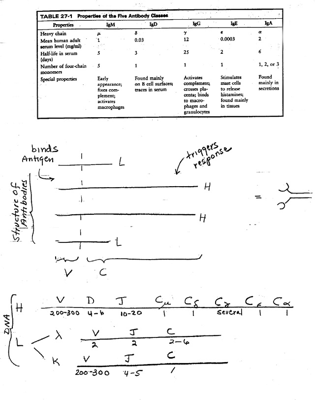
1. There are 5 main classes of Antibody -- IgM, IgD, IgG, IgE, and IgA. See table on handout 25B & Purves Table 18.3 (19.2).
2. V & C: Each Ab or Ig is made up of a V section ("variable" region or Vee) & a C section ("constant" region or Cee).
3. Variable region
a. V is specific for Ag. Determines what Ag will be bound = grabbers.
b. V is variable due to differences in sequence, not just differences in folding around Ag.
c. Every Ab or Immunogloblin (Ig) has (at least) 2 grabbers.
d. All grabbers in one Ab are the same.
e. All the antibodies made by one Ab-producing cell have the same V. All the antibodies made by descendents of that cell have very similar V's. (Minor differences are due to somatic mutation; see advanced texts if you are interested. We will ignore somatic mutation for the rest of this discussion, and assume all the antibodies made by the descendents of one cell have the same variable region.)
4. Constant region
a. C determines biological effects -- localization of Ab, and what will happen as consequence of binding Ag. (Whether complement will be activated, whether Ab will be found primarily in blood or secretions, etc.)
b. 5 main types of C regions, therefore 5 main classes of antibody. (For properties of the dif. classes see handout 25B or Purves Table 18.3 (19.2) )
c. The same V's can go with different C's. (Called "Class Switching")
(1). All the antibodies made by one Ab-producing cell do not necessarily have the same C.
(2).The antibodies made by descendents of a single cell may have different C's. The same variable region can go with different constant regions as B cell clone expands. How is this possible? Need a closer look at Ig structure
B. H vs L. See handout 25B or Purves 18.10 (19.11)
1. Every Ig has 2 kinds of chains, L ("light") and H ("heavy"). Light and heavy refer to relative differences in mol. wt.
2. Basic unit is 2 of each for a total of 4 chains. (For number of basic four-chain units per Ig, see table.)
3. Variable region (grabber) made of parts of each.
4. Each chain has a constant region
a. 2 kinds for L (kappa or lambda)
b. 5 basic kinds for H (mu, delta, gamma, epsilon or alpha)
c. Hc (constant part of H) determines class (IgM, IgD, IgG, IgE, IgA)
d. Class (determined by Hc) determines location & other aspects of function (see "special properties" in table)
d. Class switching involves the H chains only, not the L chains.
5. Myelomas & Hybridomas: Ig structure was figured out by studying proteins made by myeloma cells (cancers derived from Ab-producing cells) or hybridomas (hybrids of Ab-producing normal cells and cancer cells). Only way to get large numbers of cells all making the same Ab/Ig. See texts for significance of hybridomas and monoclonal antibodies. (Purves 18.12 (19.13))
C. Classes of Ab and class switching during development of immune response
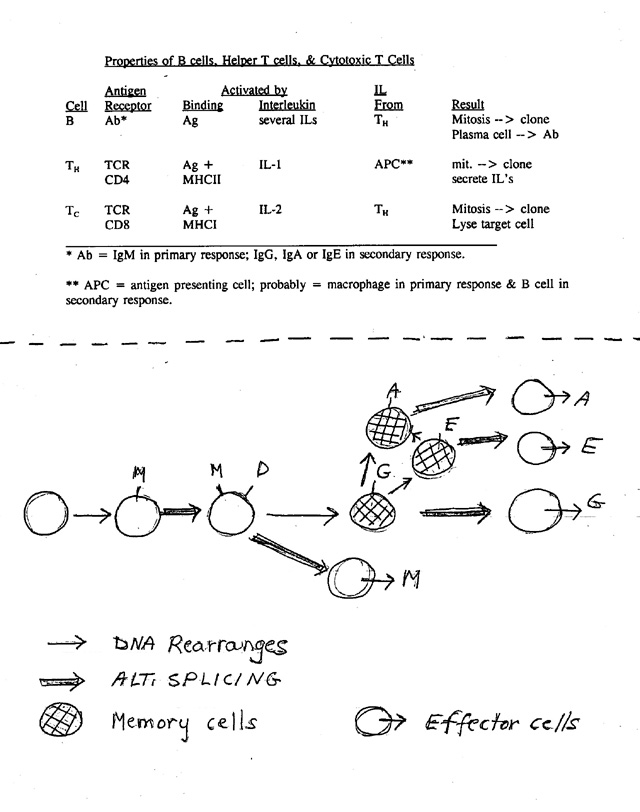
1. Order of events (see handout 25A, bottom) during immune response
a. First make M, then M + D -- all on surface.
b. Meet Ag → primary response: secrete M.
c. Meet Ag a second time → secondary response; secretes usually G but can be E or A.
d. All these Ig's combine with same Ag
2. Implications of structure and switching
a. Can make different variable regions -- zillions of them, one for each dif. epitope. So the IgG, for example, in a person is a mixture -- all IgG molecules have the same constant regions but have different variable regions.
b. During differ. stages of immune response, can make Ab with same variable region but different constant region (for H). Can switch class and/or secreted vs. surface. How is this possible?
3. What we already know: How switch from membrane bound to secreted works. Variable part stays same; Hc changes from hydrophobic to hydrophilic by alt. splicing/poly A addition.
4. What we don't know so far: How do you make so many dif. variable regions AND What changes when you switch classes (from IgM to IgG? M to M + D)? Must be rearrangement of DNA or alternate splicing of RNA. Different solutions at different steps.
Try Problems 13-1 to 13-3.
V. Structure of the DNA -- Basis of Generation of Diversity (G.O.D) and Class Switching
A. Basic idea: genes for H and L are mosaic -- Each "Gene" has several parts. See texts or handout 25B or Purves 18.18 (19.19.)
B. How "gene" is divided -- region coding for each chain (H or L) has parts coding for each type of constant region and several parts coding for the variable region .
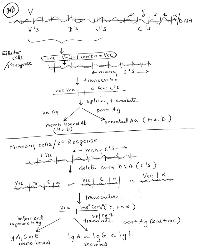
C. How DNA is used to make different antibodies (With different V's) -- DNA is rearranged -- See Handout 25C.
1. Pre Ag
a. Rearrange V/D/J region of DNA to make one coding region for variable part of H chain per naive/virgin B.
b. A similar process of DNA rearrangement occurs in DNA coding for variable part of L chain.
c. Net: Only one H chain gene and one L chain gene are rearranged and used.
2. Post Ag -- Somatic Mutation → minor changes in region of DNA coding for V regions of H & L chains. (No change in DNA coding for C regions ). In the secondary response, there is a second round of clonal selection for B cell variants making 'better Ab' -- Ab that binds Ag better (higher affinity Ab). This is why Ab made in secondary response is better at binding Ag than primary Ab. (
3. Reminder: Switching at DNA level is unique to immune system.
Note: We are going to ignore the effects of somatic mutation, but it is included here for reference.
D. Summary of G.O.D (generator of diversity) -- how get so many V's?
1. H & L mix and match -- any H chain can go with any L chain
2. Mosaic V genes -- V parts (V, D, J) of DNA coding for each chain mix and match
3. Joins are inexact -- bases can be added when you rearrange the DNA -- when join V to D etc.
4. Somatic mutation -- post Ag
E. TCR is similar, except no somatic mutation
F. Class Switching -- How DNA is used to make different versions of the same antibody (with different C regions) See Handout 25C.
1. Definition of Class switching -- cell makes antibody with same variable region and different constant region.
2. Mechanism -- Switching occurs at DNA and RNA levels.
3. Pre Ag -- M vs D
Alternate splicing allows cell to produce M and D antibodies with same V/D/J (but different constant region of H chain). For details see Purves 18-19 (19-20).
3. Post Ag -- Alt splice of RNA and/or further rearrangement of DNA → new mRNA → new version of antibody with same variable region. Can have either of the following:
a. Rearrangement (usually deletion) of DNA → gene for H chain with original variable region and a new "constant" region. Make new class of antibody. (See Purves 18.20 (19.21))
b. Alternative splicing → mRNA for secreted version of cell surface antibody -- Same H chain except it's missing part that anchors the protein in the plasma membrane. Go from making "BCR" to making secreted antibody.
Try recitation problem 14-3 & problem 13-13.
VI. Evolutionary Aspects (FYI)
A. Clonal vs. Natural Selection. Note how clonal selection and natural selection compare. In both cases, need to have many variants (diff. antibodies or dif. organisms) to be able to respond to unpredictable environmental challenges. How is this done? In both cases, make many variants and conditions select (promote propagation of) cells making the few suitable Ab (or carrying out a rare, useful function); the rest are wasted. Random generation of variants seems wasteful, but is the biological solution to preparing for change without conscious planning ahead.
B. The Major Proteins of the Immune System are Related
1. The immune system uses 3 types of proteins that have a common evolutionary origin. These are antibodies, TCR and MHC. For additional pictures see Purves 18.10 (19.11) for antibodies & Purves 18.13 (19.14) for TCR. Here are links to parallel pictures (from Alberts) of the two types of MHC, a TCR, and an immunoglobulin (showing the domains).
2. All 3 types of proteins have a "constant" part and a "variable part."
a. Constant part determines where protein is (cell surface? What kind of cell? etc.) and its general function.
b. All 3 proteins bind epitopes -- Variable part determines what antigen/epitope will bind to the protein.
3. All 3 proteins include one or more copies of the immunoglobulin domain -- a section of the protein that is similar in structure and function. this is a common theme -- the same domains are found over and over in different proteins. (Examples are SH2 domains; DNA binding domains, etc.)
4. Variable part of antibodies and TCR's are generated by rearranging the DNA; the variable part of MHC's is encoded in the germ line -- the DNA inherited in the zygote is the DNA used to code for the MHC's. However the genes for MHC's are polymorphic (have many different common alleles).
VII. Summary of Major Players in the Immune System:
| Cells | B cells, TC cells, TH cells, phagocytic cells, APC's |
| Secreted Proteins | Antibodies (Ab or immunoglobulins; 5 classes), Perforin, Cytokines |
| Cell Surface Proteins | MHC, BCR, TCR, CD4, CD8 |
The chart above summarizes the major players in immunology. You should be able to describe what each item is, its significance, and how it is related to all the others. "Secreted proteins" refers to those made by B and T cells. Proteins involved in the immune response (such as complement) that are not made by lymphocytes are not listed. See ans. to problem 13-6, table above, and the table in lecture 24 (IV-C).