C2006/F2402 '10 -- Outline for Lecture 21
(c) 2010 Deborah Mowshowitz . Last updated 04/12/2010 11:00 AM.
Handouts: 21A -- Structure
of Antibodies and their respective genes (posted version is labeled 24D)
21B --
Stages of Immune Response (Bottom 1/2 on line = bottom 1/2 of class handout)
& Rearrangement of H chain DNA (top of handout; not on line)
21C --
Lining of the GI Tract & Typical Circuit
21D (Homeostasis)
-- Seesaw view for Glucose and Temperature Regulation;
PART 1 -- WRAP UP OF IMMUNOLOGY
Problems to do on immunology (prob. set 13) will be assigned after Exam #3. Any terms in the problem book that need clarification will be explained then.
I. The Question: How do cells make so many different antibodies, MHC molecules and TCRs?
A. For MHC -- the answer is straightforward -- there are multiple genes for MHC, and each gene has many different alleles. See PP slides 32-35 of previous lecture.
B. For Antibodies & TCRs -- answer is different, & more complex. (See handouts for today or Slides 36 to end.)
1. DNA coding for antibodies & TCRs must be rearranged in each B or T cell.
2. We'll concentrate on antibodies, since the process is similar for TCRs.
3. To get the answer, we have to take a closer look at the structure of antibodies.
II. Ab Structure -- See handout 21A, picture of an immunoglobulin (from Alberts), or Sadava fig. 18.9 (18.10). What is the molecular structure of antibody molecules? Why do we need to know?
A. V vs C -- types of Immunoglobulin (Ig).
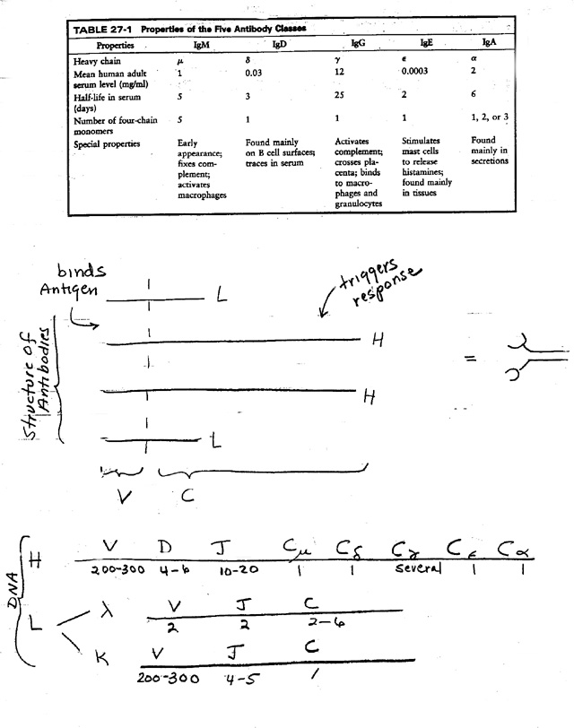
1. There are 5 main classes of Antibody -- IgM, IgD, IgG, IgE, and IgA. See table on handout 21A & Sadava Table 18.3. (Also PP slide 40 of previous lecture.)
2. V & C: Each Ab or Ig is made up of a V section ("variable" region or Vee) & a C section ("constant" region or Cee).
3. Variable region
a. V is specific for Ag (or epitope). Determines what Ag will be bound = grabbers.
b. V is variable due to differences in sequence, not just differences in folding around Ag.
c. Every Ab or Immunogloblin (Ig) has (at least) 2 grabbers.
d. All grabbers in one Ab are the same.
e. All the antibodies made by one Ab-producing cell have the same V. All the antibodies made by descendents of that cell have very similar V's. (Minor differences are due to somatic mutation; see advanced texts if you are interested. We will ignore somatic mutation for the rest of this discussion, and assume all the antibodies made by the descendents of one cell have the same variable region.)
f. DNA coding for V region is rearranged during development -- therefore a huge number of different V sequences are possible. Details below.
4. Constant region
a. C determines biological effects -- localization of Ab, and what will happen as consequence of binding Ag. (Whether complement will be activated, whether Ab will be found primarily in blood or secretions, etc.)
b. 5 main types of C regions (in the DNA), therefore 5 main classes of antibody. (For properties of the dif. classes see handout 21A or Sadava Table 18.3 )
c. The same V's can go with different C's. (Called "Class Switching")
(1). All the antibodies made by one Ab-producing cell do not necessarily have the same C.
(2).The antibodies made by descendents of a single cell may have different C's. The same variable region can go with different constant regions as B cell clone expands. How is this possible? Need a closer look at Ig structure (& structure of the coding DNA).
B. H vs L. See handout 21A or Sadava fig. 18.9 (18.10). Also PP slide 41.
1. Every Ig has 2 kinds of chains, L ("light") and H ("heavy"). Light and heavy refer to relative differences in mol. wt.
2. Basic unit is 2 of each for a total of 4 chains. (For number of basic four-chain units per Ig, see table.)
3. Variable region (grabber) made of parts of each. Both L and H chains have variable regions.
4. Each chain has a constant region
a. 2 kinds of constant region for L (kappa or lambda)
b. 5 basic kinds of constant region for H (mu, delta, gamma, epsilon or alpha)
c. Hc (constant part of H) determines class (IgM, IgD, IgG, IgE, IgA)
d. Class (determined by Hc) determines location & other aspects of function (see "special properties" in table)
e. Class switching involves the H chains only, not the L chains.
f. Mechanism of class switching: DNA rearrangements or alternative splicing of RNA are required for cells to make an H chain with the same old variable region, but a new constant region. (Details below.)
5. Myelomas & Hybridomas: Ig structure was figured out by studying proteins made by myeloma cells (cancers derived from Ab-producing cells) or hybridomas (hybrids of Ab-producing normal cells and cancer cells). Only way to get large numbers of cells all making the same Ab/Ig. See texts for significance of hybridomas and monoclonal antibodies. (Sadava 18.11 (18.12))
C. Classes of Ab and class switching during development of immune response
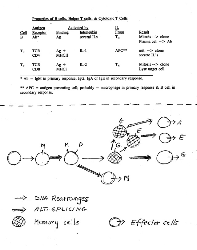
→ primary response: secrete M.1. Order of events during immune response as B cell matures (see handout 21B, bottom)
a. First make M, then M + D -- all on surface.
b. Meet Ag
c. Meet Ag a second time → secondary response; secretes usually G but can be E or A.
d. All these Ig's combine with same Ag -- only constant region of H chain switches.
2. Implications of structure and switching
a. Can make different variable regions -- zillions of them, one for each dif. epitope. So the IgG, for example, in a person is a mixture -- all IgG molecules have the same constant regions but have different variable regions.
b. During different stages of immune response, cells can make Ab with same variable region but different constant region (for H).
(1). Can switch class of antibody
(2). For any particular class, can switch from membrane bound Ab on surface to secreted Ab.
c. How does switching of class and/or location of Ab occur?
3. What we already know: How switch from membrane bound to secreted works. Variable part stays same; Hc changes from hydrophobic to hydrophilic by alt. splicing/poly A addition. (See handout 9B &/or PP slides 46 & 47 from previous lecture.)
4. What we don't know so far:
a. How do you make so many dif. variable regions? Requires rearrangement of germ-line DNA.
b. What changes when you switch classes (from IgM to IgG? M to M + D)?
a. Requires rearrangement of DNA or alternate splicing of RNA.
b. Different solutions at different steps.
Try Problems 13-1 to 13-3.
III. Structure of the DNA coding for Antibodies -- Basis of Generation of Diversity (G.O.D) and Class Switching
A. Basic idea: genes for H and L are mosaic -- Each "Gene" has several parts. See texts or handout 21A or Sadava 18.16 (18.18.)
B. How "gene" is divided -- region coding for each chain (H or L) has parts coding for each type of constant region and several parts coding for the variable region .
C. How DNA is used to make different antibodies (With different V's) -- DNA is rearranged -- see handout 21B, top, step 1.
1. Pre Ag
a. Rearrange V/D/J region of DNA to make one coding region for variable part of H chain per naive/virgin B. Put one V next to one D next to one J. Delete all the intervening segments.
b. A similar process of DNA rearrangement occurs in DNA coding for variable part of L chain.
c. Net: Only one H chain allele and one L chain allele are rearranged and used. Therefore each cell makes only one type of variable region.
2. FYI: Post Ag -- Somatic Mutation** → minor changes in region of DNA coding for V regions of H & L chains. (No change in DNA coding for C regions ). In the secondary response, there is a second round of clonal selection for B cell variants making 'better Ab' -- Ab that binds Ag better (higher affinity Ab). This is why Ab made in secondary response is better at binding Ag than primary Ab.
3. Reminder: Switching at DNA level is unique to immune system. Parts of genes for antibodies (or TCRs) can be rearranged using enzymes that cut DNA and join (recombine) DNA segments that were not contiguous in the germline DNA. Some of these enzymes are restricted to certain immune cells. (For details see PP slides 38-49 of Lecture 20.)
**Note: We are going to ignore the effects of somatic mutation, but it is included here for reference.
D. Summary of G.O.D (generator of diversity) -- how get so many V's?
1. H & L mix and match -- any H chain can go with any L chain
2. Mosaic V genes -- V parts (V, D, J) of DNA coding for each chain mix and match
3. Joins are inexact -- bases can be added when you rearrange the DNA -- when join V to D etc.
4. Somatic mutation -- post Ag
E. TCR genes are similar, except no somatic mutation. Genes are mosaic, and are rearranged. The proteins that are made have more than one chain; each TCR has constant and variable regions. See TCR picture from Alberts.
F. MHC genes are different. Reminder: no DNA rearrangements are involved in expression of MHC genes. MHC genes in all cells of a person are the same -- same as in germ line. (But unrelated people almost always have different MHC alleles.)
G. Class Switching -- How DNA is used to make different versions of the same antibody (with different C regions). See handout 21B (top).
1. Definition of Class switching -- cell makes antibody with same variable region and different constant region.
2. Mechanism -- Switching occurs at DNA and RNA levels.
3. Pre Ag -- M vs D
Alternate splicing allows cell to produce M and D antibodies with same V/D/J (but different constant region of H chain). For details see Sadava fig. 18.17 (18.19).
3. Post Ag -- Alt splice of RNA and/or further rearrangement of DNA → new mRNA → new version of antibody with same variable region. Can have either of the following:
a. Rearrangement (usually deletion) of DNA → gene for H chain with original variable region and a new "constant" region. Make new class of antibody. (See Sadava fig. 18.18 (18.20)). See 21B, top, step 2.
b. Alternative splicing → mRNA for secreted version of cell surface antibody -- Same H chain except it's missing part that anchors the protein in the plasma membrane. Go from making "BCR" to making secreted antibody.
Try recitation problem 13-3 & problem 13-13 (Ignore IL-2 receptors).
IV. Evolutionary Aspects
A. Clonal vs. Natural Selection. Note how clonal selection and natural selection compare. In both cases, need to have many variants (diff. antibodies or dif. organisms) to be able to respond to unpredictable environmental challenges. How is this done? In both cases, make many variants and conditions select (promote propagation of) cells making the few suitable Ab (or carrying out a rare, useful function); the rest are wasted. Random generation of variants seems wasteful, but is the biological solution to preparing for change without conscious planning ahead.
B. The Major Proteins of the Immune System are Related
1. The immune system uses (at least) 3 types of proteins that have a common evolutionary origin. These are antibodies, TCR and MHC. For additional pictures see Sadava fig. 18.9 (18.10) for antibodies & Sadava fig. 18.12 (18.13) for TCR. Here are links to parallel pictures (from Alberts) of the two types of MHC, a TCR, and an immunoglobulin (showing the domains).
2. All 3 types of proteins have a "constant" part and a "variable part."
a. Constant part determines where protein is (cell surface? What kind of cell? etc.) and its general function.
b. All 3 proteins bind epitopes -- Variable part determines what antigen/epitope will bind to the protein.
3. All 3 proteins include one or more copies of the immunoglobulin domain -- a section of the protein that is similar in structure and function. this is a common theme -- the same domains are found over and over in different proteins. (Examples are SH2 domains; DNA binding domains, etc.)
4. Variable part of antibodies and TCR's are generated by rearranging the DNA; the variable part of MHC's is encoded in the germ line -- the DNA inherited in the zygote is the DNA used to code for the MHC's. The DNA from MHC is NOT rearranged. However the genes for MHC's are polymorphic (have many different common alleles).
V.
Summary of Major Players in the Immune System:
| Cells | B cells, TC cells, TH cells, phagocytic cells, APC's (Antigen presenting cells) |
| Secreted Proteins | Antibodies (Ab or immunoglobulins; 5 classes), Perforin#*, Cytokines* or Interleukins##* |
| Cell Surface Proteins | MHCI, MHCII, BCR, TCR, CD4, CD8 |
The chart above summarizes the major players in immunology. You should be able to describe what each item is, its significance, and how it is related to all the others. "Secreted proteins" refers to those made by B and T cells. Proteins involved in the immune response (such as complement) that are not made by lymphocytes are not listed. See ans. to problem 13-6.
*Terms with a star have not been discussed in detail, but you should be aware of their general roles. (See notes below.) You will not be asked any questions on the final about perforin, somatic mutation, cytokines, or ILs. Some of these terms are used in the problem book in order to provide more thorough explanations.
Notes:
#Cytotoxic T cells use proteins called perforins to make holes in their targets. Then other proteins enter the holes and trigger apoptosis. Note complement is similar to perforins but works on prokaryotic invaders; perforins work on rogue eukaryotic cells. Many texts say perforin lyses cells -- it makes holes in membrane, and then water enters, causing cells to swell and burst. (This is the way complement kills bacteria.) Newer data indicates perforin works to trigger apoptosis.
##Cytokines are paracrines that affect cells of the immune system. ILs (interleukins) are cytokines that are secreted by WBCs. Many steps in the the adaptive immune response are dependent on binding of the proper IL to its receptor. The two terms, cytokine and IL, are often used interchangeably.
PART II -- Homeostasis
I. Introduction to Physiology & Multicellular organisms
A. Single cell Life Style vs. Multicellular
1. Single celled organisms
a. Surrounded by external environment -- Can't change or regulate it
b. Have one basic function -- grow and multiply
c. Respond to external conditions (since can't change them) to maintain optimal intracellular state
(1). Pick up and/or dump what is necessary for metabolism
(2). Keep intracellular conditions (pH, level of amino acids, oxygen, etc.) as constant as possible and expend minimal energy by adjusting rates of transcription, enzyme activity, etc.
d. Note no specialization: each cell does all possible functions
2. Multicellular organisms & Homeostasis
a. Each cell in organism surrounded by internal environment. Extracellular fluid (ECF) that makes up internal environment is composed of:
plasma = liquid part of blood = fluid between blood cells
interstitial fluid (IF) = fluid between all other cells
b. Organism as whole can regulate composition of internal environment (milieu); therefore can maintain relatively constant external environment for each cell. Process of maintaining a relatively constant internal environment (of whole organism) = homeostasis.
c. Each cell has two basic functions
(1). Grow or maintain itself as above
(2). Specialized role in maintaining homeostasis of whole organism
d. Cells are Specialized. Maintenance of homeostasis requires co-operation of many different cell types, not just circuits within a single cell.
Summary of Above:
| Unicellular Organisms | Multicellular Organisms | |
| What surrounds cell? | External environment | Internal environment of organism |
| Can organism regulate what surrounds each cell? | No | Yes |
| How many functions of each cell? | 1 | 2 or more |
| Is cell specialized? | No | Yes |
B. Organization -- How are cells set up to co-operate in a multicellular organism? See 21C.
1. Cells, Tissues & the 4 major tissue types (5, if you count the blood separately) -- see lecture #4, & Sadava 40.7 (41.2)
2. Organs
a. Made of (different kinds of) tissues.
b. Example: lining of GI tract. Has layers of different tissues -- epithelial, connective, muscle, and nervous; these serve primarily for absorption (of material from lumen), support, contraction, and regulation respectively. (See handout 21C or Sadava fig. 40.7 (41.2)) The blood (a type of connective) doesn't really fit in this classification -- serves for transport of materials in and out.
3. Systems -- Group of Organs → body or organ system. Work together to maintain homeostasis for some component. See Sadava 40.1 (41.1). Number of systems depends on who's counting. Usual # is 8-12; see Sadava Table 41.1 (in 7th ed only) for a list.
a. Immune system -- responds primarily to internal changes caused by presence of foreign organisms (or their macromolecules) -- responds to viruses, bacteria, cancer cells, etc. (Graft rejection, allergy, etc. are side effects of this.)
b. Other systems -- respond to changes in internal mileu caused by other factors.
III. How
is a component of the internal milieu regulated?
A. General Principle -- Homeostasis is maintained by Negative Feedback
1. What is negative feedback? The system is self correcting -- it responds so as to decrease deviations from the set point. Deviations in either direction (too high or too low) are corrected back to standard (the set point).
2. How is negative feedback different from positive feedback? In positive feedback, the system responds so as to increase deviations from the set point -- a small deviation triggers a bigger one, which triggers a bigger one and so on. The deviations get bigger and bigger until → boom! (Contractions leading to birth, summary of graded potentials until you generate an AP, lactation, etc.)
3. Results of negative FB: Value of regulated variable (blood glucose, or temperature) does not remain exactly constant, but stays within narrow limits.
4. Note on Terminology Some of the terms discussed here are used differently in molecular biology and in physiology. Fortunately, the meaning is usually obvious from the context. For example, the terms "effector" and "negative feedback" are used differently in the two contexts.
In physiology, negative and positive feedback are defined as above. For negative fb, it doesn't matter whether the corrections are achieved by inhibition (turning off the heater; stopping glucose production) or acceleration (turning on the air conditioner, increasing glucose uptake from blood).
In biochemistry, negative feedback usually means inhibition of an earlier step (& positive fb usually means activation).
'Effector' is also used differently in physio and biochem; see below.
B. Example #1 -- Regulation of blood glucose levels. The see-saw view. See handout 21D or Sadava fig. 50.19.
1. Have a regulated variable -- glucose level in blood.
2. Need a sensor (or receptor) -- to measure levels of "regulated variable" (glucose). Here, sensor is in pancreas.
3. Need effector(s) -- to control levels of regulated variable (glucose) -- usually have one or more effectors that respond in opposing ways. In this case, effectors for uptake of glucose are liver, adipose tissue, and skeletal muscle; effector for release of glucose is liver.
Note on terminology: In physiology, "effector" usually means "a tissue or organ (like muscle or liver) that carries out an action and thus produces an effect." In this example, the effectors = organs that act to raise or lower the blood glucose. In molecular biology, the term "effector" is usually used to mean "a modulator of protein function." A modulator = a small molecule (like an inducer, enzyme activator etc.) that binds to a protein, alters the shape and/or function of the protein, and thus triggers an effect.
4. Have a set point -- the level the regulated variable (blood glucose) should be. Set point is also sometimes used to mean the level at which corrections (to raise or lower the value) kick in.
In most cases, there is no significant difference between these two definitions of set point. In some cases, the desired value (first definition) and the value at which corrections occur (second definition) may be different. For example, there may be two cut-off points-- upper and lower, that bracket the desired level of a regulated variable. At levels above or below the respective cut-off points, messages are sent to the appropriate effectors to take corrective action. The term "critical values" is sometimes used instead of "set points" to describe the cut-off point(s).
5. Signaling -- need some signal system to connect the sensor(s) and the effector(s). Can be nervous &/or hormonal. In this case, primary (but not only) signal is hormonal & primary hormones (signals) are insulin & glucagon.
6. Operation of Negative Feedback -- the system responds to negate deviations from the set point. Important features:
a. Works to stabilize levels of blood glucose (the regulated variable)
b. System is self-correcting -- Deviations in either direction (if blood glucose is either too high or too low) are corrected.
c. There are two opposing actions by effectors, not just one.
(1). If [G] gets too high, effectors take G up from blood. (top half of seesaw diagram)
(2). If blood [G] gets too low, effector releases G to blood. (bottom half of seesaw diagram)
d. Negative feedback is not always inhibition. The deviation from the set point may be fixed by accelerating, not inhibiting, a process. In negative feedback, deviations from the set point can be corrected either by speeding up a process (such as glucose uptake) or slowing down a process (such as glycogen breakdown to glucose). For example:
(1). If blood [G] goes up, uptake from blood increases and glycogen breakdown decreases. In this case, an increase in glucose uptake is used to help decrease the deviation from the set point.
(2). If blood [G] falls, release into blood increases, and glycogen synthesis decreases. In this case, an increase in glucose release is used to help decrease the deviation from the set point.
(3). In both cases, one process is increased and another is inhibited to help decrease the deviation from the set point.
7. Net Result -- regulated variable ([G] in blood) is not constant, but stays close to set point.
See problem 5-1 & 5-2 a & b.
C. Example #2 -- Regulation of body temperature (in humans) -- the see-saw view (handout 21D)
1. Note many features are same as in glucose case. (Can you list them??)
2. Features not found in glucose case:
a. Multiple sensors in different places (for core and skin temp.). How to integrate multiple inputs?
b. Nature of Signal -- Signals are neuronal, not hormonal
c. Integrative center (IC)
(1). Role of IC: Compares set-point to actual value, sends appropriate message to effectors.
(2). Type of IC
(a). Sensor/IC function may be combined, as in Glucose example.
(b). Separate IC needed if there are multiple sensors, as in this case. IC co-ordinates incoming information from multiple sensors
(3). In this example, IC = hypothalamus (HT)
3. Organs/body systems involved as effectors
|
Effector |
Action To Raise Temp |
Action To Lower Temp |
|
Skeletal muscles |
Contraction generates heat (shivering) |
None |
|
Smooth muscle of peripheral blood vessels in skin |
Muscles contract; vessels constrict to reduce heat loss |
Muscles relax; vessels dilate to increase heat loss |
|
Sweat glands |
None |
Produce sweat; evaporation increases heat loss |
|
Brain |
Behavioral (nonphysiological) responses-- put on coat, curl up, etc. |
Behavioral (nonphysiological) responses -- take off coat, etc. |
4. Cooling vs. Heating -- What can effectors do? Effectors can increase or decrease heat loss; can only increase heat generation. (Cannot decrease heat generation.) Therefore ability of humans to cope with very cold environments is better than their ability to cope with excessively hot environments.
5. Does drinking make you warmer in winter? See this 'Really?' column from the NYTimes.
Try Problem 5-2, c. & 5-5.
C. Body Temperature and the General Case -- The Circuit View -- handout 21C, top.
1. Circuit = 1 loop of seesaw. Seesaw = double circuit. Often two circuits to make opposite types of corrections.
2. Signals: Signals can be hormonal or neuronal.
3. Afferent vs Efferent Signals. Bottom half of circuit has two arms -- afferent vs efferent
→ toward effectors
Afferent information goes from sensors → in to IC
Efferent goes out of IC
4. Regulation vs Control.
a. Regulation/regulated variable: The variable (glucose level) you wish to keep at an approximately constant level is said to be "regulated."
b. Control/controlled process: The processes that alter levels of the regulated variable (glucose uptake, release or shivering, sweating, etc.) are said to be "controlled."
c. What's the difference?
The point of the system is to maintain homeostasis of blood glucose levels, internal temperature, etc. Not to maintain homeostasis of rates of glucose uptake, sweating, etc.
The value of the regulated variable stays about the same; the rates of the controlled processes (glucose uptake, heat loss, heat generation, etc. ) can vary as much as necessary to achieve homeostasis of blood glucose levels or temperature.
5. May be multiple effectors and/or sensors.
6. IC (when there are multiple inputs) is nervous tissue or brain.
a. Major Role -- Compares current value to set point; sends appropriate message to effectors.
b. Adjustments -- IC can adjust set points and/or critical points. Why bother? Fevers & feedforward:
(1). Fevers -- Raise set point for body temperature and critical points for shivering/sweating
Shivering and sweating both kick in at higher temps. (You don't have to cool off as much to start shivering and you need to heat up more to start sweating.) Raises set point (desired level) & actual level of internal body temperature.
Why fevers? High temperatures prevent bacteria from obtaining iron from host & improve immune function.
(2). Feedforward or anticipation -- Planning ahead. Altering set points and/or critical points to adjust to anticipated factors. (Or you can think of it as just ignoring the usual critical points.) Examples:
Body temperature: Skin temperature affects critical temperature/set points for generating heat and/or shivering. If body is cold, but it's warm outside, shivering can be postponed, saving energy, and you'll still warm up. This is equivalent to lowering (or ignoring) set point/critical points for shivering, not changing set point of internal body temperature. Changes what effectors and what controlled processes you use to warm up, but not the end result.
Secreting insulin when you start to digest food in the stomach, but before the digestion products (glucose, amino acids etc.) reach the blood. This way tissues will be ready to take up the glucose as soon as it enters the blood.
D
. What other components of internal milieu are regulated besides glucose, temperature? Many nutrients like amino acids; concentrations of water, salts and ions (Na+, K+ etc.), gases (CO2, O2), waste products, volume & pressure of blood, and pH.Try Problems 5-3, 5-4 & 5-9
A & B, & 5-10.