C2006/F2402 '10 OUTLINE OF LECTURE #22
(c) 2010 Dr. Deborah Mowshowitz, Columbia University, New York, NY. Last update 04/21/2010 07:11 PM .
Handouts: 22A. Endocrine vs Exocrine Glands & catecholamines,
22B (Hormones
Overall)
This topic is not covered in Becker. It is covered in Sadava/Purves. If you want a
more detailed treatment, any physiology book will do. There are lots of good
physiology books available; the one by Sherwood & the one by
Silverthorn have been used here for the last few years. There is an endocrinology book on line through Pubmed. Go to
books to see the list of books available or to search by topic. Also
don't forget about
Kimball's
biology pages.
I. Intro to Homeostasis, cont.
A. Regulation of Body Temperature (handout 21D) -- See notes of last time.
B. The Circuit View (handout 21C) -- See notes of last time.
II. Matching circuits and signaling -- an example: How the glucose circuit works at molecular/signaling level
Re-consider the circuit or seesaw diagram for homeostatic control of blood glucose levels -- what happens in the boxes on 21D? It may help to refer to the table below.
A. How do Effectors Take Up Glucose?
1. Major Effectors: Liver, skeletal muscle, adipose tissue
2. Overall: In response to insulin, effectors increase both uptake & utilization of glucose. Insulin triggers one or more of the following in the effectors:
a. Causes direct increase of glucose uptake by membrane transporters
b. Increases breakdown of glucose to provide energy
c. Increases conversion of glucose to 'stores'
(1). Glucose is converted to storage forms (fat, glycogen), AND
(2). Breakdown of storage fuel molecules (stores) is inhibited.
d. Causes indirect increase of glucose uptake by increasing phosphorylation of glucose to G-P, trapping it inside cells
3. How does Insulin Work?
a. Receptor: Insulin works through a special type of cell surface receptor, a tyrosine kinase linked receptor; See Sadava 15.6. Insulin has many affects on cells and the mechanism of signal transduction is complex (activating multiple pathways). In many ways, insulin acts more like a typical growth factor than like a typical endocrine. (Insulin has GF-like effects on other cells; is in same family as ILGFs = insulin like growth factors). More on GFs and TK receptors later.
b. How Does Insulin Increase Glucose Uptake in different effectors?
(1). In resting skeletal muscle & adipose tissue -- mobilizes GLUT 4: In these tissues insulin mobilizes transporter for facilitated diffusion (of glucose) -- GLUT 4 protein -- promotes fusion of vesicles containing the transporters with plasma membrane. No other hormone can cause this effect.
(2). In liver: Liver (& brain) can take up glucose without insulin -- they do not use GLUT 4. They use different transporters (GLUT 1, 2 &/or 3) located permanently in the plasma membrane.
(a). In liver: Insulin promotes glucose uptake in liver, but not directly. Insulin promotes uptake by increasing phosphorylation (trapping) and utilization of glucose.
(b). Note: Insulin has no affect on glucose uptake in brain.
(3). Working skeletal muscle: Insulin is not required for uptake of glucose in working skeletal muscle because exercise mobilizes GLUT4 in skeletal muscle. (Another good reason to exercise.)
c. Other Effects: In many tissues, insulin promotes utilization of glucose:
(1). Activates appropriate enzymes for synthesis of storage forms of metabolites -- synthesis of glycogen, fat, and/or protein.
(2). Inhibits enzymes for breakdown of stores.
(3). Can promote utilization (breakdown) of glucose for energy.
d. Significance: Some effects of insulin are mimicked by other hormones, but mobilization of GLUT4 cannot be triggered by any other hormone. Therefore loss of insulin, or lack of response to insulin, is very serious, and causes diabetes type I or II, respectively. (See absorptive state, below.)
B. How do Effectors Release Glucose?
1. Primary Effector for Release = Liver
a. Only organ that can release significant amounts of glucose into blood -- why? Liver has phosphatase for G-6-P. Muscle and adipose tissue don't.
b. Other tissues can breakdown stores (fat, glycogen) to release fatty acids or lactate into blood, but cannot release glucose.
2. Overall: Stores are broken down to generate small molecules; liver releases glucose into blood.
3. Role of Glucagon
a. Receptor: Glucagon works through a G protein linked receptor that triggers the cAMP pathway (as for epinephrine). Therefore it activates PKA; see text or handout on glycogen metabolism for details.
b. Effects: Primary physiological effect is on liver; generally promotes production/release of glucose, not uptake or utilization. (Glucose is produced both by breakdown of glycogen, and build up from lactate = gluconeogenesis. See texts if you are interested in details of gluconeogenesis.)
c. Receptor triggers same pathway as epinephrine. Note that the same signaling pathway can be used for two different hormones (epinephrine & glucagon).
(1). Epi.(epinephrine) & glucagon bind to different receptors, but both receptors activate the same G protein and trigger the same series of events → cAMP → etc. so can get same response to both hormones in same tissue (if both receptors are present).
(2). Two hormones control same process (glycogen metabolism) for different purposes -- Epi to respond to stress; glucagon to respond to low blood sugar (maintain homeostasis).
(3). Different tissues can respond differently to these hormones. How? Both hormones trigger production of cAMP and activation of PKA. But there are differences in which receptors are present and/or which targets of PKA:
(a). Receptors: Receptors present on cell surface determine which tissues will respond to each hormone.
Muscle has Epi receptors (but no glucagon receptors); therefore responds to Epi but not glucagon
Liver has receptors for both epi and glucagon and responds to both.
(b). Targets: Even if receptors are same, different enzymes and/or processes are available to be affected by same kinase. For example, glycogen metabolism in liver vs. skeletal muscle. Both tissues break down glycogen in response to epi, but result is different.
In muscle, breakdown to lactate, and release lactate to blood.
In liver, breakdown to glucose - P, and release glucose into blood.
d. Significance: Actions of glucagon can be mimicked by other hormones; there is no known medical condition caused by lack of glucagon. (See post absorptive state below.)
C. Overall Function of Effectors -- Summary:
1. Liver -- both releases glucose to blood and stores excess (as glycogen).
a. Carries out both storage and release of glucose so acts as buffer.
b. Only organ that can release significant glucose into blood (kidney may do some).
c. Takes up glucose without insulin -- uses GLUT 2 (always in plasma membrane), not GLUT 4. Insulin stimulates phosphorylation & utilization of glucose, not direct uptake.
2. Muscle -- stores or releases energy.
Takes up glucose; stores excess as glycogen.
When glycogen is broken down, releases lactate, not glucose, into blood.
3. Adipose Tissue -- stores or releases fat/ fatty acids.
Uses up glucose & fatty acids; stores excess as fat.
When fat is broken down, releases fatty acids into the blood.
4. All three organs co-operate -- for example lactate generated in muscle is not broken down further in muscle -- it is shipped to liver and metabolized further in the liver. For many more details than you need see Sadava 50-20 (7th ed only) or advanced texts.
D. Absorptive vs Postabsorptive State -- A more complex view of the circuit
1. What is really being regulated by insulin & glucagon? Really two different things:
a. Maintenance of glucose homeostasis
b. Managing an episodic event (eating) -- this can be considered just another example of homeostasis -- here the 'episodic' nature of eating generates two basic states that must be controlled differently to maintain homeostasis.
2. There are two main states of food (not just glucose) supply. A detailed diagram of fuel traffic in both states (that goes way beyond what you need) is in Sadava fig. 50.20 (7th ed only) and in all physiology books.
a. Absorptive -- anabolic → synthesis & storage of macromolecules; glucose is primary energy source. In this state, right after you eat, the risk is that blood glucose levels will rise too much. Absorptive state is completely dependent on insulin. Insulin affects all three effector organs.
b. Postabsorptive -- catabolic → breakdown of macromolecules to release glucose*; fatty acids are primary energy source (except in brain). In this state, between meals, the risk is that blood glucose levels will fall too much. Postabsorptive state is largely caused by lack of insulin; also utilizes glucagon, but stress hormones (cortisol and epinephrine) can fill in for glucagon. Glucagon mainly affects liver.
*(Gluconeogenesis also occurs in liver = resynthesis of glucose from smaller molecules; see texts if you are interested.)
For questions on this topic
see problem set 7,
questions 7-23 to 7-26, and 4R-3.
To review and to be sure you have this topic straight, fill in the
following tables:
| Responds to Insulin? | Responds to Glucagon? | Can Release Glucose to Blood? | Uses GLUT 4 | Can take up Glucose w/o Insulin? | |
| Skeletal Muscle | + | - | - | + | only when working; not at rest |
| Liver | |||||
| Adipose tissue |
* |
||||
| Brain | - | - | - | - | + |
* Adipose tissue has glucagon receptors, but there is no known response to physiological levels of glucagon.
| Insulin | Glucagon | |
| Type of Receptor/signaling pathway | ||
| Effect on blood glucose -- release or uptake? | ||
| Effect on glycogen -- synthesis or breakdown? | ||
| Result of intracellular glucose metabolism -- use it up or generate it? | ||
| Mobilize GLUT4? | ||
| Effect on pathways of intracellular glucose production -- inhibit or stimulate? |
III.
Introduction to Hormones (Endocrines) & Growth Factors
A. How to describe or classify hormones?
1. Many Possible Classification Schemes -- Hormones can be classified by effect, chemical nature, source (which gland?), target cells, etc. etc. See Topic V (for reference) for a extensive list.
2. Today: We will look at (1) processes controlled by hormones, (2) the major hormone producing glands, (3) details for specific hormones.
B. Summary of typical hormone roles and examples. See Becker Table 14-3 or Sadava fig. 41.5 (table 42.1) for a list of hormones by type of function (Becker) or by source (Sadava).
1. Stress response -- cortisol, epinephrine. Regulate heart rate, blood pressure, inflammation, etc.
2. Maintenance of Homeostasis -- insulin, glucagon. Regulate blood glucose/energy supplies and concentrations of substances in general. Maintain more or less constant conditions = homeostasis.
3. Regulation of episodic or cyclic events -- estrogen, insulin, oxytocin -- regulate lactation, pregnancy, effects of eating, etc.
4. Growth/overall regulation -- growth factors, tropic hormones -- regulate production of other hormones. (Note: not all GF's are endocrines.)
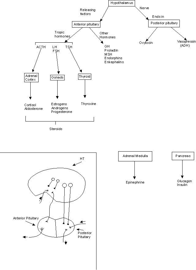
C. Overview of Major Glands & Hormones -- see handout 22B for overview. For a complete list see Sadava fig. 41.5 (Table 42.1)
1. Pancreas
a. endocrine gland -- secretes glucagon and insulin into blood -- These control blood sugar balance, as explained last time.
b. also exocrine gland -- secretes digestive enzymes through duct to GI tract.
What's the difference between endocrine & exocrine glands? See handout 22A.
(1). Exocrine Gland
When gland forms, epithelial layer leaves duct to outside.
Secretion from gland flows into duct → outside or lumen.
Examples:
(i) sweat, mammary & tear glands → secretion → outside
(ii) stomach glands & pancreas → secretion → lumen.(2). Endocrine Gland
When gland forms, epithelial layer pinches off leaving no duct
Secretion (hormone) from gland enters blood.
Example: gonads, pancreas, adrenal.
(3) Both types get precursors for secretions from blood
c. Regulation -- secretion of glucagon/insulin controlled by blood sugar levels and by input from sympathetic (in response to stress).
2. Adrenal Medulla & Cortex See Sadava fig. 41.11 (42.10).
a. Medulla (nervous)
(1). Stimulated by nerves
(2). Derived from neural tissue; part of autonomic NS.
(3). Secretes compounds that can act as transmitters (when signal cell to cell) but act as hormones (neuroendocrines) here -- are released into the blood. Note same compound can act as a transmitter or a neuroendocrine.
(4). Major hormone = epinephrine (adrenaline); also secretes some norepinephrine (noradrenaline)For structures see handout 22A. More details below.
(5). Receptors. Receptors for these hormones/transmitters are same adrenergic receptors (α & β) discussed previously.
b. Cortex (epithelial)
(1). Stimulated by a hormone (ACTH)
(2). Derived from epithelial tissue
(3). Produces steroids = corticosteroids. For structures see Sadava fig. 41.12 (42.11).
(4). Part of HT/AP axis; more details below or next time.
3. Additional info on dopamine (DA) & related compounds = catecholamines
a. Structures: See handout 22A for structures of catecholamines = epinephrine (aka adrenaline), norepinephrine (aka noradrenaline), and dopamine. These are all modified amino acids derived from tyrosine. All water soluble. (Note thyroxine is also derived from tyrosine but is not a catecholamine; it is lipid soluble -- see below.)
b. Receptors: There are multiple receptors for all of the catecholamines. Receptors are classified by their ligands and response to drugs.
(1). Dopamine has its own receptors, separate from the adrenergic receptors (for epi and norepi).
(2). FYI: All adrenergic receptors bind to both epi and norepi. Some receptor types bind better to (have higher affinity for) one, some to the other, some equally well to both.
Epinephrine acts mostly through beta adrenergic receptors.
Norepinephrine mostly through alpha adrenergic receptors.
c. Mechanism of action: All receptors for all catecholamines are G protein linked; effects of hormones on any particular cell type depend on
(i) what receptors are present
(ii) what G protein each receptor activates. Each G protein does one (or more) of the following:
activate adenyl cyclase
inhibit adenyl cyclase
activate phospholipase C.
See Previous lectures & problem 6-21 and 6-22 for examples of different responses to epi due to diff. receptors. For an example of the effects of dopamine, see problem 6-24.
4. Hypothalamus (HT) -- neuroendocrine. HT is IC (integrative center) for many homeostatic circuits.
a. Inputs: 3 types of inputs
(1). neuronal
(2). hormonal
(3). local conditions. HT has sensors for some variables such as temperature, osmolarity.)
b. Outputs: To pituitary (also called hypophysis)
(1). To anterior pituitary (AP); also called adenohypophysis
(2). To posterior pituitary (PP); also called neurohypophysis
c. Details of structure and HT hormones below. (For structure, see handout 22B.)
5. Post. Pit. (Sadava fig. 41.6 (42.5).
a. Hormones = ADH = antidiuretic hormone (aka vasopressin) and oxytocin.
(1). ADH. Affects (primarily) water retention; has 2 names because discovered twice from different effects. Details of action to be described when we get to kidney. (Works through IP3 or cAMP.)
(2). Oxytocin. Affects milk ejection, uterine contractions -- works (at least in part) through IP3 to affect Ca++ and therefore contraction
b. Origin/action of hormones: Peptides are very similar in structure (homologous = share common evolutionary origin) but bind to different (G protein linked) receptors → dif. effects.
6. Anterior Pit -- Hypothalamus (HT) / Pituitary Axis
a. HT/Ant. Pit -- 3 stages
(1). HT → hormones (releasing factors) that signal the AP. Hormones go direct to AP through portal vessel (see handout 22B).
(2). AP (anterior pituitary) → tropic hormones (ACTH, LH, etc.) that signal to glands (endocrine tissue)
(3). Glands → lipid soluble hormones (steroids & TH) which control their target organs. Overall:
HT
→ releasing hormone → AP → tropic hormone → TARGET GLAND → hormone → TARGET TISSUE → action.b. Example: How HT controls adrenal cortex
HT
→ CRH → AP → ACTH → ADRENAL CORTEX → corticosteroids → TARGET TISSUES → action(1). HT secretes corticotropin releasing hormone (CRH)
(2.) Ant. Pit responds by secreting ACTH (adrenal cortex tropic hormone; also called adrenocorticotropin) into general circulation
(3). Adrenal cortex produces three major types of steroids = corticosteroids. For structures see Sadava 42.12 (41.11) .
(a). Glucocorticoids. Ex: cortisol -- involved in long term stress response (after epinephrine wears off) -- has multiple effects/targets, for ex. suppresses immune system. ACTH controls production of cortisol.
(b). Mineralocorticoids. Ex: aldosterone -- regulates salt balance (to be discussed when do kidney). Major control of aldosterone production is by other factors, not ACTH. ACTH has only a weak effect on aldosterone.
(c). Sex Steroids -- cortex produces low levels of sex hormones (both androgens and estrogens) in both sexes post puberty. That's how females get 'male' hormones and vice versa.
c. AP also → "other hormones" (GH, Prolactin, etc.) that signal to nonendocrine tissues.
7. There are other glands/hormones -- the list so far is not exhaustive but covers most of the major players. See texts for complete lists.
It is worthwhile to memorize most of handout 22B in order to keep all the hormones and glands straight.
IV. Details of HT& Pituitary Set Up
A.
Structure -- Two parts of pituitary (AP and PP) develop and function separately; connected differently to HT.1. Ant. Pit. (epithelial)
a. Blood vessel goes direct from HT to AP. See Sadava fig. 41.7 (42.7); handout next time.
Normally, blood flows from artery in general circulation → some tissue → vein in general circulation. Blood does not normally go direct from one organ to another.
Direct (Portal) vessel connects 2 organs.
b. Release: Hormones released from HT into the blood travel through portal vessel direct to AP.
c. AP is Epithelial. AP consists of epithelial tissue that grows up from mouth.
2. Post. Pit. (nervous)
a. Cells connect HT & PP. Some cells of HT have bodies in HT and axons/terminals in posterior pituitary. (Sadava fig. 41.6 (42.5).
b. Release: Hormones (neuroendocrines) are released from nerve endings (terminals) in post. pit → general blood supply.
c. PP is neural. PP consists of neural tissue that grows down from brain.
B. Hypothalamic Hormones
1. Outputs (to AP): Some cells in HT release hormones from HT itself. (As vs. cells that connect to post. pit.)
a. Release hormones into portal vessel (see above) that goes direct to anterior pituitary.
b. Hormones are release factors. Hormones released by HT affect production/release of other hormones by ant. pit.
c. Affect on release -- 'release factors' can be stimulatory (RH's such as ACTH-releasing hormone) or inhibitory (IH's such as prolactin release-inhibiting hormone = PIH)
d. All HT hormones (except PIH = dopamine) are peptides/proteins.
2
. Outputs (to PP): Some cells in HT release hormones (ADH & oxytocin) from nerve endings in PP. Hormones are peptides; made in cell body, packaged in vesicles, vesicles travel down MT's to end of neurons, hormones released by exocytosis.V. How
to Keep Track of Hormones
-- How to Classify Hormones & Growth Factors
(or Signal Molecules in General). The following is meant as a check list to help
you keep track of the various signal molecules. It is for reference & study
purposes; it will not be discussed in class.
Some of these questions/categories overlap, and you can't answer all
the questions for all the hormones, growth factors, etc., but the list helps to organize the information you do
have.
1. Type of Action -- Is it paracrine, endocrine (hormone), growth factor, neurotransmitter, etc.? (See handout 12A)
2. Chemical nature -- Is it a peptide, amino acid or derivative, fatty acid or derivative, or steroid? See Becker table 14-4.
3. Where is it made? In what gland or tissue? (HT? pancreas?) See Sadava fig. 41.5 (Table 42.1)
4. Target Cells -- where does it act? (Muscle and liver? Just liver?)
5. Mechanism of signal transduction
A. Location/type of receptor on target cells -- Is receptor on surface or intracellular? TK* or G protein linked?
B. Type of signal transduction -- Is there a second messenger? Which one? If none, what links receptor to intracellular events?
C. Intracellular mode of action -- what mechanism is used to get the end result? Is there a change in enzyme activity? change in transcription? both? change in state of ion channel?
6. What actually gets done? What happens?
A. Biochemically speaking: Which target enzymes, proteins or genes are affected (glycogen phosphorylase activated? Gene for enzyme X transcribed?)
B. Physiological End Result: Another hormone secreted? Glycogen broken down, & Glucose in blood up? Note the "result" may have several steps, and more than one can sometimes be considered "the end."
C. What's the (teleological) point? What overall function is served by the signal molecule's action?
1. One list of possibilities: Homeostasis, response to stress, growth*, maintenance of some cycle;
2. An alternative version of the list: Regulation of rates of processes, growth & specialization, Conc. of substances, and response to stress.
3. The 2 lists are really the same = homeostasis (control of rates & concentrations), response to stress, & regulation of growth (unidirectional and cyclic).
* Details of TK linked receptors have not been discussed (yet) in 2010. The point so far is that they are not GPCRs, and work differently.
Next Time: Details of HT/AP axis, examples of circuits using hormones & nerves, and signaling with TK receptors.