Electrostatic Properties of Semiconductor Nanocrystals
Single-molecule Surface Enhanced Raman Spectroscopy
Optical and Electronic Properties of Carbon Nanotubes
Iron Oxide Nanocrystal Synthesis and Reactions
Physical Properties of Arene Liquid Crystals
Electronic Properties of Pentacene
Electrostatic Properties of Semiconductor Nanocrystals
Oksana Cherniavskaya, Chaya Ben-Porat
Semiconductor nanocrystals have attracted much attention over the last
decade due to their unique physical properties, and
potential use for a wide range of applications ranging from
all optical switching to biological labels. While the
electronic and vibrational properties of semiconductor
nanocrystals have been studied extensively, the
electrostatic properties have received little attention. A
complete understanding of the electrostatic properties of a
single semiconductor nanocrystal is not only of fundamental
interest, but is also required to fully understand a
nanocrystal's optical, electronic, and vibrational
properties.
Using an atomic force
microscope, we study directly the electrostatic properties
of single CdSde, PbSe, and gold nanocrystals. Previously, the electrostatic
polarization of semiconductor nanocrystals has been either
indirectly measured or inferred, but not measured directly.
We now have the ability to measure electrostatic properties
directlly, and in real time. In particular, we use a
technique called electrostatic force microscopy (EFM) to
measure the dielectric constant and electrostatic
polarization of single nanocrystals in dry air at room
temperature. We also combine this technique with optical
methods to study the properties of a phtotexcited
nanocrystal. Here are images of CdSe nanocrystals using
EFM.
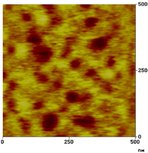
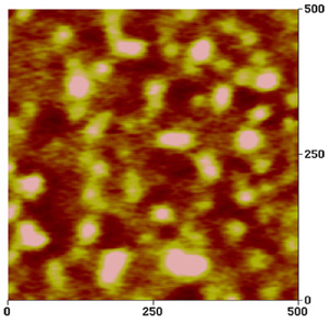
Fig. EFM image of CdSe
nanocrystals on the PVB/HOPG substrate taken by Todd Krauss. (a) Tapping mode
AFM height image. The scale in the vertical direction is
10 nm. The images in (b) and (c) correspond to the charge
and dielectric constant of the particles on the
surface.
Single-molecule Surface Enhanced Raman Scattering
Ken Bosnick, Jiang Jiang, Mathieu Maillard
It has been known for some time now that silver surfaces with nanoscale features enhance the Raman scattering
from molecules adsorbed on them. This effect is termed "Surface Enhanced Raman Scattering" (SERS). Recently, extreme
examples have been found, where the enhancement reaches levels permitting single-molecule detection. We study one
such system, namely Rhodamine 6G on optically resolvable aggregates of silver nanocrystals, with the goal of
understanding the physics that underlies this phenomenon.
Our first study involves exploring the relationship between the local electromagnetic field enhancement and
the large SERS enhancement by measuring both resonant Rayleigh scattering spectra and Rhodamine 6G Raman spectra
from single Ag particle aggregates. Our apparatus combines the techniques of dark-field optical microscopy for
resonant Rayleigh measurements and grazing incidence Raman spectroscopy. The Rayleigh spectra show that the
citrate-reduced Ag colloid is extremely heterogeneous. Only the larger particles, in part created by salt
induced aggregation, show a large SERS effect. In agreement with the work of Nie and Emory, we find that a few
nanocrystals show huge single molecule R6G SERS intensities. While all SERS active particles have some resonant
Rayleigh scattering at the 514.5 nm laser wavelength, we find that there is no correlation between the resonant
Rayleigh spectra and the SERS intensity.
Atomic force microscopy (AFM) measurements show that the Ag nanoparticles that yield surface-enhanced Raman
scattering (SERS) of single molecules of Rhodamine (R6G) are all compact aggregates consisting of a minimum of two
individual particles. Comparison of 514.5 and 632.8 nm excitation shows that the single molecule R6G signal is
significantly higher when the excitation wavelength is resonant with the absorption band of R6G and suggests that
the Raman excitation spectrum follows the absorption profile for R6G. We have also observed an interesting superlinear
power dependence of the SERS signal. On average, by increasing the incident power by 2 orders of magnitude and
decreasing the integration time by the same factor to maintain constant fluence, increases of 4 to 6 times were
observed in the SERS intensity. We explain these results in terms of model where the R6G molecule that yields
single molecule SERS signals is located at the junction of two Ag nanoparticles. Classical electromagnetic field
calculation also shows that just the enhanced electric field at nanoparticle junctions could give a SERS enhancement
of ten orders of magnitude.
The single-molecule SERS signal fluctuates and blinks on a second time scale, presumably due to movement of the molecule
in to and out of the SERS active junctions. We studied the depolarization of the signal and found it to be independent
of the fluctuations and Raman shift, to vary from one spot to the next, and to be highly dependent on the direction of
the incident electric field. The signal is dominated by a continuum emission, which is assumed to arise from electronic
Raman processes in the metal that are triggered by chemisorption of the molecule in the hot spot. The source of
the Raman signal is therefore highly localized around the hot spot. The local structure determines the depolarization
properties and is often extremely anisotropic. Conversely, the Rayleigh scattering does not depend on the molecule or
the hot spot and is delocalized over the whole aggregate, which is always found to be highly isotropic. The Rayleigh
scattering therefore does not fluctuate and is always highly polarized.
Optical and Electronic Properties of Carbon Nanotubes
Xiadong Cui, Matthew Sfeir, Gordana Dukovic
A variety of optical methods are used to nonivasively probe how the chemical
and physical environment alters the electronic and optical properties of single wall carbon nanutbes. These methods include
dark field optical microscopy, Raman microscopy, extinction, and fluorescence experiments. Small variation in chirality
and diameter results in a large
variation of physical properties. These optical measurements, combined with transport and EFM studies in collaboration
with IBM work towards understanding nanotube characteristics on an individual level.
Iron Oxide Nanocrystal Synthesis and Reactions
Jing Tang
Magnetite (Fe3O4) and maghemite (γ-Fe2O3) are widespread in the environment, despite the fact that both
are thermodynamically unstable with respect to hematite (α-Fe2O3) in the presence of oxygen. They are found in bacteria
and insects, weathered soils and clays, rocks, natural-atmospheric and polluted aerosols, and even on the surface of
the Mars. Near room temperature magnetite very slowly oxidizes to maghemite, and then at higher temperatures, to
hematite. The oxidation of magnetite to maghemite is thus a significant environmental process, and we study the optical
spectroscopy and oxidation kinetics of aqueous colloidal nanocrystals. Magnetite and maghemite nanoparticles are
also widely used as ferrofluids such as in rotary shaft sealing, dynamic loudspeakers, and computer hard drives,
and also have medical use in, for example, magnetic resonant imaging and targeted drug transportation.
Most prior oxidation studies have been done on larger dry magnetite particles in air, using X-ray diffraction
and/or chemical analysis methods. Oxidation is believed to occur through the outward diffusion of iron cations.
Presumably at the surface the Fe reacts with O2 and forms a thin layer of epitaxial maghemite. We study the oxidation
of magnetite nanoparticles to maghemite in aqueous solution via the loss of near IR absorption. The kinetics do not
fit simple rate-laws, but rather fit the diffusion in a sphere model used for dry oxidation.
The Fe diffusion constants and the activation energy in water agree well with the values reported for dry oxidation
of larger particles. In addition to the thermal oxidation, photo effects on the oxidation were also studied.
Physical Properties of Arene Liquid Crystals
Quyen Nguyen
Measurable polar order is a highly desirable attribute that is responsible for many useful properties such as piezo-, pyro-,
and ferroelectricity. Creating spontaneously polar materials on nanometer length scales is an important challenge that
requires finding alternatives to the poling fields used to align the dipoles of bulk materials. This study utilizes atomic force microscopy and electrostatic force microscopy to investigate the
orientation of overcrowded aromatics in films with submonolayer coverage. The results demonstrate that
the side chains in the molecules can be used as a tool to control the molecular order and orientation in thin
films. For molecules that do not self-associate well, the interaction with the substrate dominates, and the
molecules orient with their aromatic planes parallel to the surface. These monolayers have measurable
polar order.
Electronic Properties of Pentacene
Liwei Chen
Conjugated organic materials have shown great promise in novel electronic
and optoelectronic applications. Pentacene, for example, has been used as
the active material in field-effect transistors. In high-performance
organic field effect transistors, current modulation is believed to be
restricted to the top few layers of the organic molecules at the interface
with the gate dielectric. Consequently, understanding the role of surface
structure and properties on this 2-D electronic transport process is
important from both fundamental and technological perspectives.
We use a combination of local scanning probe technique and optical methods
to address these issue. In particular, physical vapor deposition has been
used to controllably grow single domain nanocrystals of pentacene on silicon
substrate with a thin oxide layer. We are currently investigating the
electrostatic characteristics of the interface of pentacene nanocrystals and
the gate oxide with electrostatic force microscopy (EFM). In collaboration
with researchers at IBM, we are also studying the transport of electrons and
holes at this interface with pulsed laser probes.
Last modified: Wednesday, 05-Feb-2003 13:11:54 EST