
Recent News from Columbia
Statement from Columbia University President Minouche Shafik
To deescalate the rancor and give us all a chance to consider next steps, I am announcing that all classes will be held virtually on Monday. During the coming days, a working group of Deans, university administrators and faculty members will try to bring this crisis to a resolution.
How Should Communities Adapt to the Changing Climate?
A new database offers communities, leaders, and researchers unprecedented access to climate data and cutting-edge models.
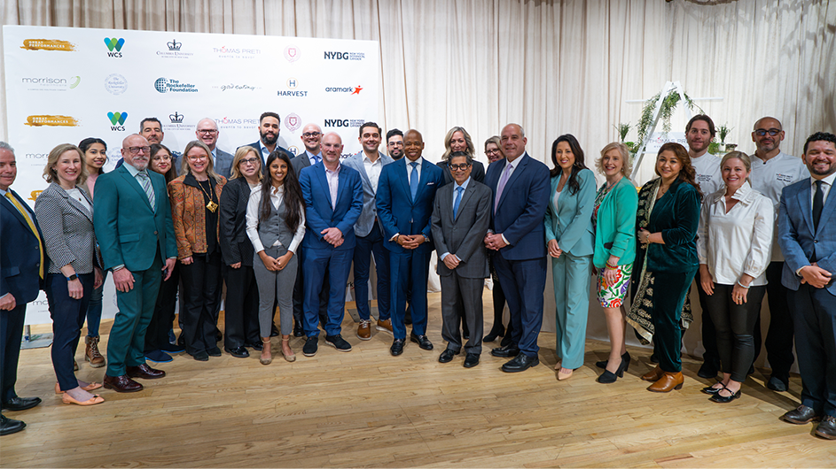
Columbia Commits to Reducing Food-Related Carbon Emissions 25% by 2030
The University joins New York City mayor's office at Plant-Powered Carbon Challenge launch.

Research & Discovery


Arts & Humanities

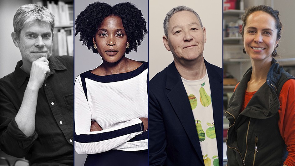
Co-writer Ann Cooper’s Newshawks in Berlin describes the perils of reporting from war zones.

The GSAPP professor’s outdoor sculptures advocate for the experimental preservation of U.S. embassies.

In his new book, Dennis Yi Tenen presents AI as a matter of collaborative labor history.
Campus & Community


National & Global Affairs


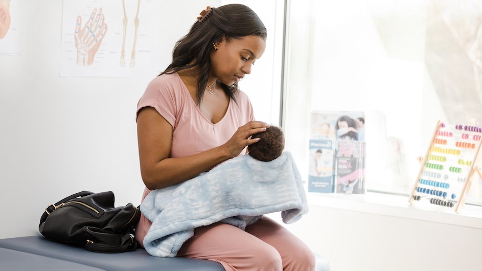
The event convened leading policymakers, scholars, and advocates to discuss a range of issues, from reproductive rights to gender equity in the workplace to technology-facilitated gender-based violence.


Columbia in the News
Los Angeles Times, April 19
NBC News, April 18
Quanta Magazine, April 16


