ENDODONTIC ACCESS AND ANATOMY
Endodontic coronal cavity preparation |
- Outline form -
- external outline form evolves from internal anatomy of the pulp as opposed to operative outline form which is based on external anatomy.
- size of pulp chamber
- larger in young teeth
- smaller in older teeth
-
varies with shape of crown
- age of tooth
- functional activity
- history of tooth
- number and curvature of root canals
- in Endodontic Access preparation convenience form regulates the ultimate outline form
- Convenience form -
- objectives of Endodontic Convenience form
1. unobstructed access to the canal orifice
2. direct access to the apical foramen - freedom within coronal cavity to reach apex in unstrained position
3. cavity expansion to accommodate filling techniques
4. complete authority over enlarging instrument- *inadequate convenience form will lead to:
1. perforation of root
2. ledging of root
3. instrument breakage
4. incorrect shape of completed canal
5. improper debridement -
Removal of remaining carious dentin -
- eliminates bacteria from interior of tooth
- eliminates discolored tooth structure
- eliminates saliva leaking into prepared access cavity
- if too much tooth structure is lost which prevents placing of rubber dam and sealing against saliva gingivoplasty or crown lengthening may be necessary. - Toilet of cavity -
- all caries, debris and necrotic material must be removed from the pulp chamber before proceeding to root preparation.
-
Intra-radicular preparation and cleaning and shaping -
- will be discussed at another lecture
CANAL MORPHOLOGY - see appendix in syllabus
- flushing the access chamber prevents:
1. obstruction with debris during canal enlargement
2. soft debris from chamber from increasing bacterial population in
canal
3. coronal debris from staining crowns especially in anterior teeth
Use of radiographs. An x-ray shows only one two dimensional view of the three dimensional tooth. In preparing outline and convenience form the operator must visualize the total three dimensional morphology of the tooth.
Maxillary incisors
- relatively straight canals
- lateral incisors may have apical curvature to labial or distal or palatal
Access:
- always on lingual surface of tooth
- large triangular funnel shaped coronal preparation
- begin with fissure bur at high speed
- perpendicular to lingual surface of tooth
- penetrate enamel
- change direction of bur so it is parallel to long axis of tooth
- before pulp chamber is entered, change to round bur at low speed.
Mandibular incisors
- least likely teeth to need endodontics
- some roots have labial or distal curvatures
- must explore for second canal by extending adequately into cingulum
- triangular shaped access
- very similar in coronal appearance
Maxillary-mandibular canine
- very stable teeth - usually last ones lost
- one large pulp cavity
- maxillary canine - one canal
- mandibular canine - 43% have 2 roots, 2 canals
- access shape - ovoid funnel shaped preparation.
Maxillary premolars
- first premolars - mostly 2 canals
- second premolars - mostly l canal
- access-ovoid shaped in bucco-lingual direction
- pulp broad bucco-lingually
- narrow mesiodistally
Buccal Object Rule (Clark's law) -
- to be used in orienting between two canals on two dimensional x-ray
- can be used on any multiple canal tooth
- the buccal object rule states that on an angled x-ray, the object (instrument
or canal) farthest from the film (most buccal) will appear projected further
from the x-ray source compared with a second object closer to the film.
- example: the buccal root will always appear distal to the lingual root
when an x-ray source is directed from the mesial toward the distal aspect.
- to remember - MBD - in Mesial angled x-rays Buccal object is projected
to the Distal
- DBM - in Distal angled x-ray Buccal object is projected to the Mesial.
MANDIBULAR PREMOLARS
first premolars
- rounded root
- narrower mesio-distal than bucco-lingual mostly one canal
second premolar
- access of first and second premolars is ovoid- shaped extending from cusp tip to cusp tip through occlussal surface
MOLARS
Maxillary First Molars
- three well separated roots
- palatal root - longest
- mesio buccal root- broad bucco-lingually
- mostly 2 canals
- distobuccal - smallest root
- straightest root
- always look for four canals in all first molars
- second mesial canal usually located in line with the groove between
the mesiobuccal and palatal canals.
Access:
- Blunted triangular outline
- base of triangle toward buccal
- apex of triangle toward palatal
- orifice positioned at each angle of the triangle
- access cavity is entirely within mesial half of the tooth
- triangular access can be extended to blunted triangle to insure locating
second mesial canals if present
- entire roof of chamber should be removed to insure proper cleaning
Maxillary Second Molar
- root formation may be different from first molar
- usually three canals
- access similar to maxillary second molar (blunted triangular - outline)
- always look for four canals
- more variability of anatomy in second and third molars compared to first
molar.
Mandibular first molar
- has two well formed roots
- mesial root - has two canals (buccal and lingual)
- distal root - has one or two canals
- always look for four canals in all first molars
- access - rhomboid/quadralateral shape of access to allow for exploration
of second distal canal
- access cavity within mesial half of tooth but extended as far distally
as necessary to allow for ease of positioning of instruments and filling
materials
Mandibular second molars
- two roots
- usually three canals
- always look for four canals (rhomboid/quadralateral access outline)
- more variability of anatomy in second and third molars as compared with
first molar
Difficulties caused by poor access preparation
1. Inadequate Opening
- compromised cleaning and shaping of canals
- compromised instrumentation
- coronal discoloration
- prevents good fillings
- inadequate extension -leaves orifice only partially exposed (mouse-hole
effect)
- instrument breakage in canal
- perforation
- ledging
2. Mutilation of coronal tooth due to removal of too much tooth structure
- coronal fracture
- mutilation of root - ledging, perforation
3. Inadequate caries removal
- carious destruction of tooth
- discoloration
4. Labial perforation
5. Furcal perforation
- difficult to repair
- can cause periodontal destruction
- weakens tooth structure - can lead to fracture
6. Misinterpretation of angulation of tooth
- common with full crown restorations
- can lead to root perforations which can cause periodontal problems
7. Entering the wrong tooth
- common problem in teeth that are identical coronally, i.e., mandibular
anteriors
- caused by placing the rubber dam clamp on the wrong tooth
8. Allowing debris to clog orifices
- dentinal debris
- amalgam fillings
IMPORTANT NOTE: The morphology described represents ideal situations. Many teeth that need root canal treatment will no longer have this ideal morphology due to loss of tooth structure, large restorations or crown restorations. By remembering to view the pulp chamber as a three dimensional object, proper access can still be obtained.
Appendix
In order to carry out endodontic treatment, it is (among other things) necessary to know the interior anatomy of the teeth. This outline gives schematic pictures of the anatomy of the fully developed permanent teeth. Also, typical access preparations of the various teeth are described.
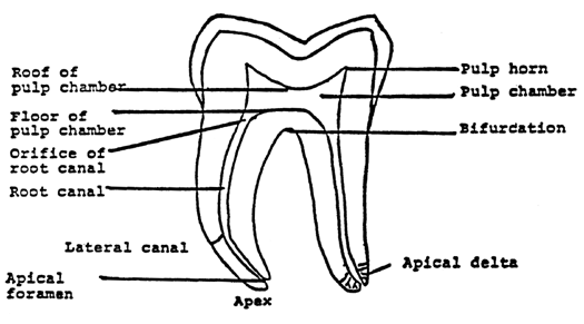
The shape of the pulp chamber is usually a diminution of the crown. The pulp horns extend towards the cusps in premolars and molars, and towards the incisal edge in incisors and canines. In the following drawings (and in most radiographs) root canals seem to be straight and the walls seem to be smooth. As a matter of fact, in each tooth there are ramifications, lateral canals and other divergencies from this seemingly straight course. The lateral canals contain periodontal tissues and they can appear everywhere in the root. They are especially prevalent in the most apical part of the root where they form apical deltas.
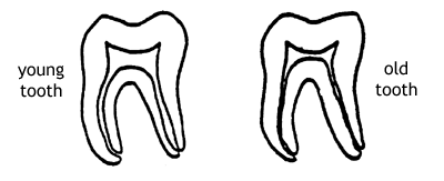
Permanent dentin production makes the pulp cavity more and more narrow as the patient grows older. Moreover, denticles and hard tissue formation adjacent to cavities also contribute to a narrowing of pulp chamber and root canals.
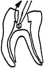
The apical foramen is usually not situated at the "tip" of the root, but 0.5 - 1.5 mm from the apex. Thus, when a radiograph shows that the tip of a root canal instrument is at the apex, the tip of the instrument is usually beyond the apical foramen (fig). When treating a tooth it is usually not possible to determine the site of the apical foramen and therefore it is recommended to instrument and fill the canal "short of the apex" to decrease the risk of over instrumentation and over filling.
Good visibility and accessibility are necessary to carry out an endodontic treatment satisfactorily. To achieve this, the access preparation must be made so that it is possible to inspect the coronal part of the pulp cavity visually and with instruments after completion of the opening of the pulp chamber. In many instances (probably most) when patients have been referred to an endodontist because a started endodontic treatment has "gone wrong", the cause is poor access preparation.
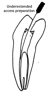
Underextended access preparations may cause canals to be overlooked, anatomical divergences will not be detected, and infected material will be left in the root canal and that necrotic tissue remaining in the pulp chamber will cause discoloration of the crown. Furthermore, a narrow access preparation may direct a bur or root canal instrument and increase the risk of perforation. It is more important to have good access than to save a cusp, because a good root filling is necessary to keep an endodontically involved tooth.
Procedure
Remove all caries and fillings that stand in the way of view or that can cause leakage. Undermined enamel shall also be removed together with parts of the crown that make accessability to the canal(s) difficult e.g., mesiobuccal cusps of molars.
 If
there is a pulp exposure, it should be widened, in order to properly determine
the extention of the pulp chamber. In this way a proper access preparation
can be made. When there is no exposure, access should be made by drilling
towards a large pulp horn or the largest area of the pulp chamber. During
the access preparation the bur should be used with a pull stroke from
the pulp chamber and out.
If
there is a pulp exposure, it should be widened, in order to properly determine
the extention of the pulp chamber. In this way a proper access preparation
can be made. When there is no exposure, access should be made by drilling
towards a large pulp horn or the largest area of the pulp chamber. During
the access preparation the bur should be used with a pull stroke from
the pulp chamber and out.
Using this technique of access preparation, it is possible to avoid perforating the floor of the pulp chamber and to get smooth walls without ledges. The access preparation is done with round burs. Long shank round burs are occasionally necessary. The use of fissure burs very often creates ledges in the floor and walls of the cavity access preparation. Such ledges make the canal instrumentation more difficult. Moreover, ledges in the dentin can diminish the tensile strength of the tooth.
When completed, the access preparation should be shaped without overhanging edges. In the following schematic drawings the access preparations are drawn with dotted lines.
Central Maxillary Incisor
|
Average
Length:
|
22.5 mm |
|
Number
of canals:
|
1 |
|
Root
development completed at 10 years of age.
|
|
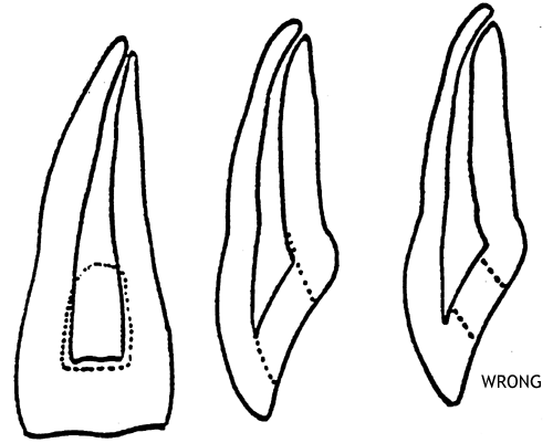
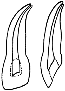
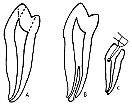
The preparation is begun from the palatal surface. The access cavity must be extended in an incisal direction. Too narrow an access cavity (according to wrong in the figure) can leave tissue remnants in the pulp horns which can cause discoloration of the crown.
Lateral Maxillary Incisor
|
Average
Length:
|
22 mm |
|
Number
of canals:
|
1 |
|
Root
development completed at 10 years of age.
|
|
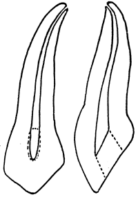
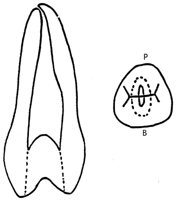
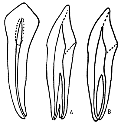
The access preparation of the lateral incisor is also begun from the palatal surface. The root canal is wide in proportion to the root and also there is usually a distopalatal curve in the apical third of the canal. Therefore, the canal must be instrumented carefully to avoid perforation.
Maxillary Canine
|
Average
Length:
|
26.5 mm |
|
Number
of canals:
|
1 |
|
Root
development completed at 14 years of age.
|
|
The access preparation is begun from the palatal surface. The root is often curved apically. This is the longest tooth and therefore considerable widening of the the root canal is needed in order to do a proper root filling.
First Maxillary Premolar
|
Average
Length:
|
20.6 mm |
|
Number
of canals:
|
1 - 19.5% |
| 2 - 79.5% | |
| 3 - 1% | |
|
Root
development completed at 13 years of age.
|
|
- When there are two root canals, one is buccal and the other is palatal.
- When there are three root canals, there are two buccal canals and one palatal.
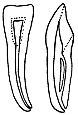
The access preparation is begun from the occlusal surface. It is sometimes necessary to cut the cusps to get an adequate view. If the access cavity is not adequately extended buccally and palatally; pulpal remnants will be left undetected. ("wrong" in the figure).
Mesially, there is a concavity of the root surface and there is an increased risk of mesio-cervical perforation during access preparation because of this.
The roots of the first maxillary premolar are often slender and curved; contours are sometimes difficult to see on the radiograph.
Second Maxillary Premolar
|
Average
Length:
|
21.5 mm |
|
Number
of canals:
|
1 - 56% |
| 2 - 42% | |
| 3 - 2% | |
|
Root
development completed at 14 years of age.
|
|
The access preparation is again made through the occlusal surface. The root canal in this usually single rooted tooth is band shaped. When there are two canals, one is buccal and one palatal. When there are three canals, two are buccal and one palatal.
First and Second Maxillary Molars
|
Average
Length:
|
first maxillary molar 20.8 mm |
| second maxillary molar 20.0 mm | |
|
Number
of canals:
|
first maxillary molar 3 - 47%, 4 - 53% |
| second maxillary molar 3 - 59%, 4 - 46% | |
|
Occasionally
there are first and second maxillary molars with 1,2, and 5 canals.
|
|
|
Root
development completed at age:
|
first maxillary molar - 9 |
| second maxillary molar - 15 | |
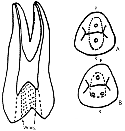
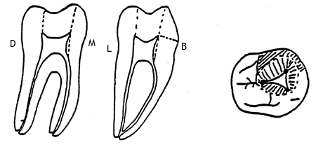
The access preparation in a maxillary molar is through the occusal surface. Very often it is necessary to reduce the mesiobuccal cusp in order to obtain straight line access to mesiobuccal canal orifice. If there are two canals, they are usually connected, but there are mesiobuccal roots that have two separate canals. Also, there are occasionally two mesiobuccal roots. The palatal and distobuccal roots have one canal each.
The mesiobuccal, distobuccal and palatal canal orifices are situated in the "Corners" of the pulp chamber. The location of these orifices represent the vertices of a triangle. The mesiopalatal orifice is mostly situated on a mentally scribed line between the mesiobuccal and palatal canal orifices (A,B). It is not uncommon, especially in the second molar, where the pulp chamber is narrow, for the canal orifices to be more or less in line. (C).
Mandibular Incisors
|
Average
Length:
|
11.7 mm |
|
Number
of canals:
|
1 - 62% |
| 2 - 38% | |
|
Root
development completed at 10 years of age.
|
|
Access preparation is done from the lingual surface of the crown. Note that the access cavity has to be extended in a linguo-cervical direction to make it possible to localize a lingual canal. Mostly, the lingual canal joins the buccal canal (see illustration), but separate foramina can occur.
Mandibular Canine
|
Average
Length:
|
25.6 mm |
|
Number
of canals:
|
1 - 57% |
| 2 - 43% | |
|
Root
development completed at 14 years of age.
Occasionally there are mandibular canines with two roots. |
|
Access preparation is done lingually. The access cavity has to be extended in a linguo-cervical direction to make a localization and instrumentation of a lingual canal possible.
The lingual canal can be situated in a lingual root (A) or join the buccal canal in a common foramen (B) or have a separate foramen within the same root as the buccal canal.
First and Second Mandibular Premolars
|
Average
Length:
|
first mandibular premolar 21.6 mm |
| second mandibular premolar 22.3 mm | |
|
Number
of canals:
|
first mandibular premolar 1 - 98%, 2 - 8% |
| (According to "clinical experience", first mandibular premolars with two root canals are more common than the frequency found by Hess in 1917.) | |
| second mandibular premolar 1 - 92%, 2 - 8% | |
|
Occasionally
there are mandibular premolars with three or more canals
|
|
|
Root
development completed at age:
|
first mandibular premolar 13 |
| second mandibular premolar 14 | |
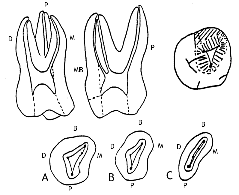
Access preparation is done occlusally. Very often the occlusal surface is pointing lingually and to make instrumentation of the canal(s) possible, the access cavity must be extended facially (according to the most facial dotted line on Fig A.) and in some cases it is necessary to reduce the facial cusp. If this is not done properly there is a risk for perforation because of interference by the facial cusp during access preparation and canal instrumentation. (C) Where there are two canals, one is buccal and the other, lingual and the division is two canals from the main canal mostly takes place in the apical third of the root (B).
First and Second Mandibular Molars
|
Average
Length:
|
first mandibular molar 21.0 mm |
| second mandibular molar 19.8 mm | |
|
Number
of canals:
|
1 - 0.5% 2 - 18.0% 3 - 79.5% 4 - 2.0% |
|
Root
development completed at age:
|
first mandibular molar 10 |
| second mandibular premolar 15 |
Access preparation is done occlusally. In most instances it is necessary to cut the mesiobuccal cusp to obtain proper accessibility.
When there is only one canal, this canal is wide, straight and centrally located. Mandibular molars with two canals have one distal and one mesial canal usually situated in distal and mesial roots. When there are three canals there are two mesial and one distal. Teeth with four canals have two mesial and two distal canals. The canal(s) of the mesial root often have many ramifications that can make their instrumentation and cleaning difficult.
|
Number of root canals (%)
|
||||
|
1
|
2
|
3
|
4
|
|
| Maxillary teeth | ||||
| Central incisors |
100
|
|||
| Lateral incisors |
100
|
|||
| Canines |
100
|
|||
| First premolars |
19.9
|
79.5
|
1
|
|
| Second premolars |
56
|
42
|
2
|
|
| First molars |
47
|
53
|
||
| Second molars |
54
|
46
|
||
| Mandibular teeth | ||||
| Central incisors |
62
|
38
|
||
| Lateral incisors |
62
|
38
|
||
| Canines |
57
|
43
|
||
| First premolars |
98
|
2
|
||
| Second premolars |
92
|
8
|
||
| First molars |
0.5
|
18
|
79.5
|
2
|
| Second molars |
0.5
|
18
|
79.5
|
2
|
| Reference Hess, W. The Anatomy of the Root Canals of the Teeth of the Permanent Dentition. 1925. |
||||
| Maxilla | Mandible | |
| Central incisors | 22.5 | 20.7 |
| Lateral incisors | 22.0 | 21.1 |
| Canines | 26.5 | 25.6 |
| First premolars | 20.6 | 21.6 |
| Second premolars | 21.5 | 22.3 |
| First molars | 20.8 | 21.0 |
| Second molars | 20.0 | 19.8 |
| Reference Black, G.V. Descriptive Anatomy of the Human Teeth, 4th Ed., Philadelphia, 1902. |
||
| Please note that these are average measures and that there is a great variation in tooth length between various teeth. | ||
| Years | |
| Central incisors | 10 |
| Lateral incisors | 10 |
| Canines | 14 |
| First premolars | 13 |
| Second premolars | 14 |
| First molars | 10 |
| Second molars | 15 |
| Reference Meyer, W. Lehrbuch der Normalen Histologie und Entwicklungs- geschichte der Zahne des Menschen. Munich. 1932. |
|