X-ray Using Parellel Cone Technique
The primary radiograph used in endodontics is the periapical radiograph. In diagnosis this film is used to identify abnormal conditions in the pulp and periradicular tissues and to determine the number of roots and canals, location of canals, and root curvatures.
For endodontic purposes the parallel technique produces the most accurate periradicular radiograph. This technique directs only the most central and parallel rays of the beam to the film and teeth, thus reducing size distortion and possibility of superimposing the zygomatic processes over the apexex of maxillary molars (top images).
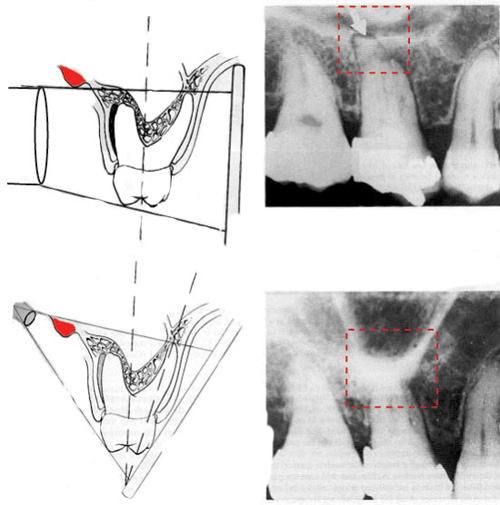
The technique produces an angle between the plane of the film and the long axis of the teeth, thus causing distortion because the tooth is not parallel to the film (bottom images).
The bisecting angle technique is not recommended for endodontic radiography; however, it may be used because of difficult anatomic configuration or patient management problem.
from Pathways of the Pulp p.89-91.