Anatomy of the Tooth
In order to carry out endodontic treatment, it is (among other things) necessary to know the interior anatomy of the teeth. This outline gives schematic pictures of the anatomy of the fully developed permanent teeth. Also, typical access preparations of the various teeth are described.
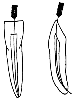
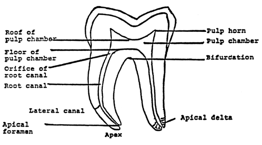
The shape of the pulp chamber is usually a diminution of the crown. The pulp horns extend towards the cusps in premolars and molars, and towards the incisal edge in incisors and canines. In the following drawings (and in most radiographs) root canals seem to be straight and the walls seem to be smooth. As a matter of fact, in each tooth there are ramifications, lateral canals and other divergencies from this seemingly straight course. The lateral canals contain periodontal tissues and they can appear everywhere in the root. They are especially prevalent in the most apical part of the root where they form apical deltas.
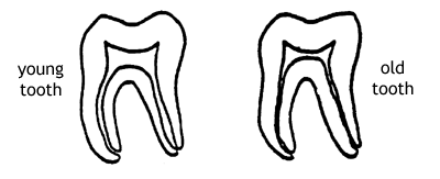
Permanent dentin production makes the pulp cavity more and more narrow as the patient grows older. Moreover, denticles and hard tissue formation adjacent to cavities also contribute to a narrowing of pulp chamber and root canals.
The apical foramen is usually not situated at the "tip" of the root, but 0.5 - 1.5 mm from the apex. Thus, when a radiograph shows that the tip of a root canal instrument is at the apex, the tip of the instrument is usually beyond the apical foramen (fig). When treating a tooth it is usually not possible to determine the site of the apical foramen and therefore it is recommended to instrument and fill the canal "short of the apex" to decrease the risk of over instrumentation and over filling.