Technical Errors 2
- Identify the triangular opacity almost in the middle of the mandible.
- Thyroid shield
- Vertebra
- Lead apron
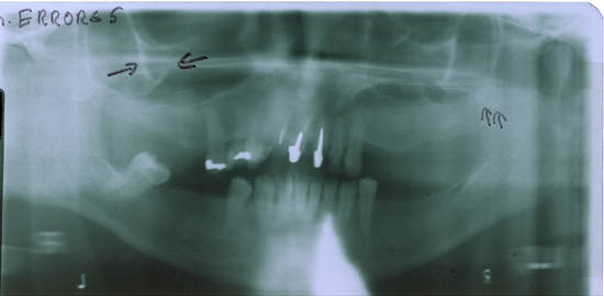
- Identify the opacity indicated by the opposing black arrows.
This is the molar bone, which is normally outside the focal trough if the patient is positioned. Note the head is elevated too much as indicated by the lack of the smile line.
- Identify the image of the anatomic structure indicated by the parallel
black arrows.
This is the outline of the lateral pterygoid plate.
- What structures attach to the lateral pterygoid plate?
The lateral ptergoid muscle to the lateral surface and the medial pterygoid muscle to the medial surface.
The outline of the lateral pterygoid plate is sometimes seen so clearly superimposed on the coronoid process that it gives the impression of a fracture of the coronoid.
One does not use a thyroid shield when taking a panoramic radiograph [Part II Board question]. The vertebra can be seen in the midline, adjacent to the opacity but not as opaque. This is the lead apron at the back of the neck. The apron must be placed as high as possible in the front of the neck to protect the bottom half of the thyroid gland but it should be low at the back of the neck to avoid the opacity seen in this pan.