SBPMD Histology Laboratory Manual
Nerve: Micrograph
Examine the electron Micrographs so that you understand the ultrastructural equivalents of the structures you have seen under the microscope.
Neuronal Cell Body | |
| Click to see enlarged view | |
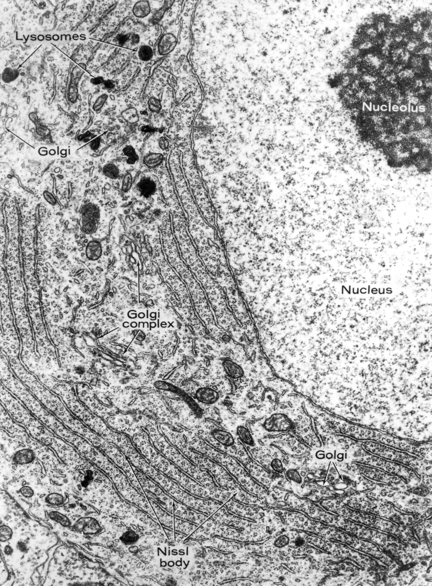 |
This portion of a neuronal perikaryon illustrates the large euchromatic nucleus with conspicuous nucleolus. There are many ribosomes, abundant rough endoplasmic reticulum (Nissl body) and Golgi. These attributes identify the cell as highly transcriptionally active. |
| |
Apical Pole of Pyramidal Neuron | |
| Click to see enlarged view | |
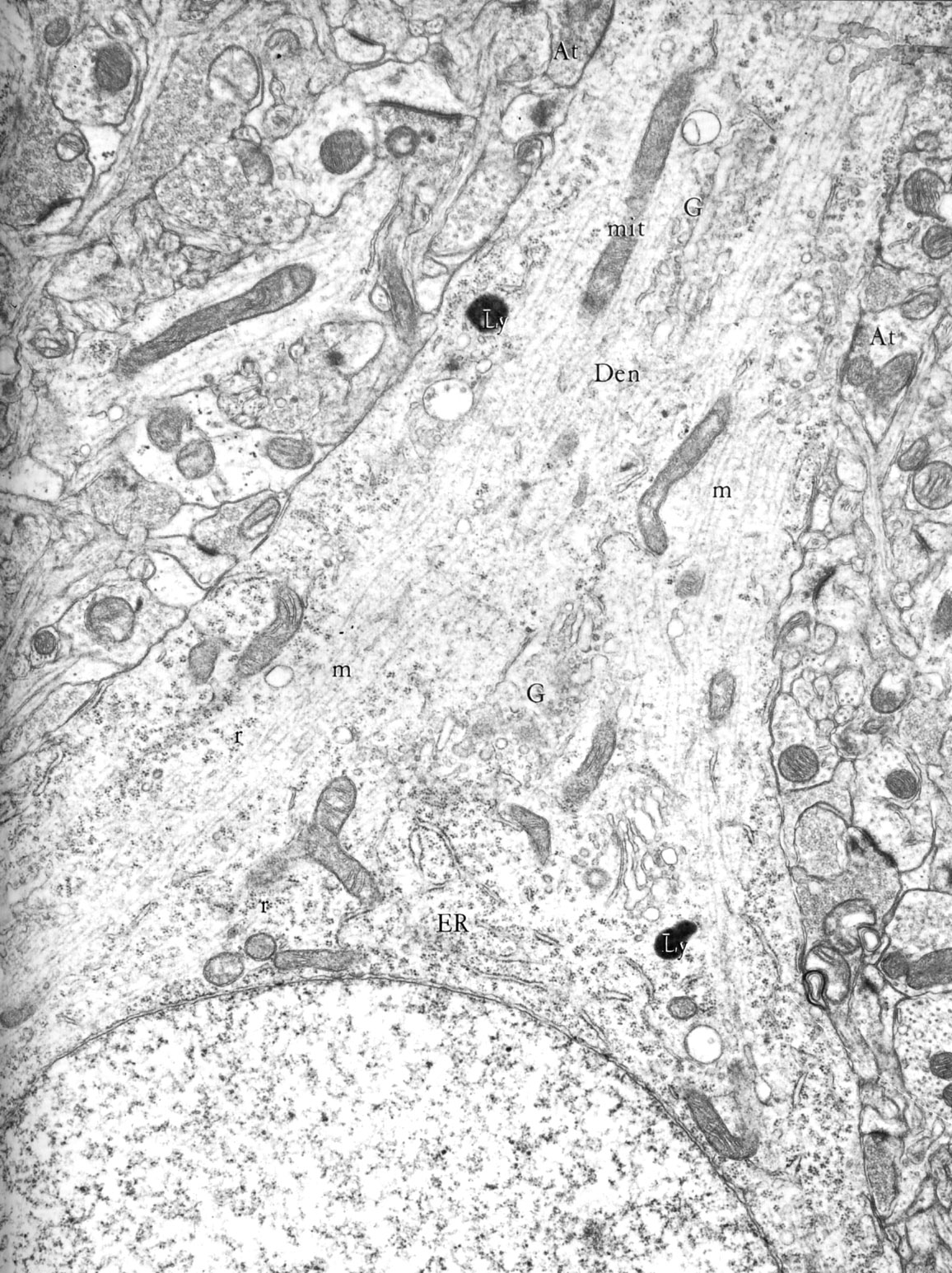 |
The nucleus of the cell is in the lower portion of the micrograph. Microtubules (m) funnel from the perikaryon into the dendrite (Den). Axon terminals (At) synapse with the neuron. There are a few cisternae of ER, some Golgi and clusters of free ribosomes. Mitochondria (mit), lysosomes (Ly). (cerebral cortex, rat) |
| |
Node of Ranvier in Peripheral Nervous System | |
| Click to see enlarged view | |
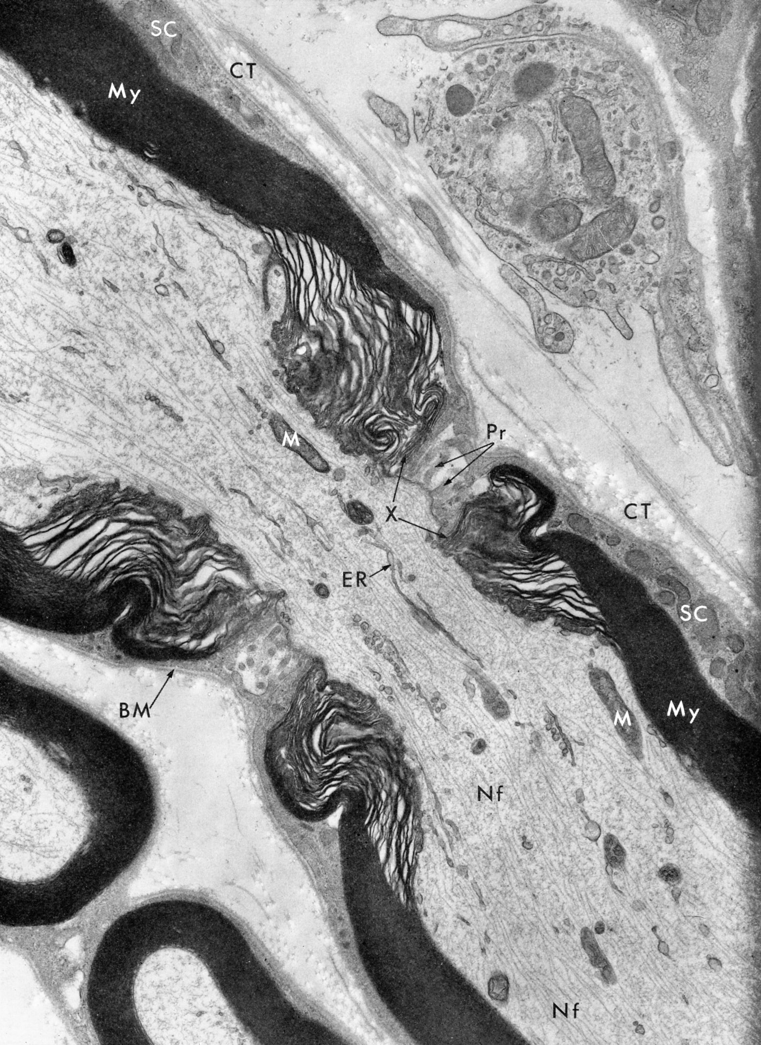 |
Nodes of Ranvier are interruptions in the myelin sheath (My) of nerves. The successive layering of the Schwann cell(Sc) membranes forms a compact tube over most of the internodal areas. Near the node, some Schwann cell cytoplasm remains in the extended margins of the sheath layers and occupies a series of liplike folds (X), which envelop the fiber. In the region of the node itself, only fingerlike process (Pr) of neighboring Schwann cells interdigitate and cover the nodal area. A basement membrane (BM) and connective tissue fibers (CT) of the endonerium complete with wrappings of the fiber. Mitochondria (M),endoplasmic reticulum (ER), neurofilaments (Nf). (sciatic nerve, mouse) |
| |
Node of Ranvier in CNS | |
| Click to see enlarged view | |
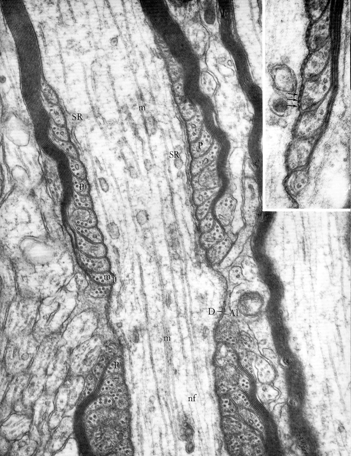 |
Node of Ranvier in central nervous system (basal ganglion, rat). At the node, the axon is bare with axolemmal densities (D). The myelin sheath becomes thinner as successive lamellae terminate to enclose pockets (P) of paranodal cytoplasm of the oligodendrocyte, which contain microtubules (m). Smooth endoplasmic reticulum (SR), neurofilaments (nf). Inset is paranodal region in optic nerve. Årrows indicate densities associated with outer leaflet of axolemma. |
| |
Axon Terminal | |
| Click to see enlarged view | |
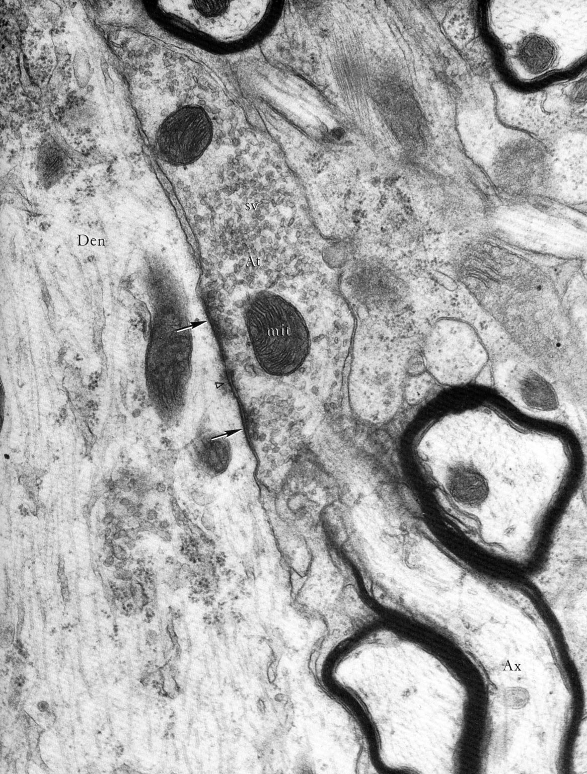 |
Axon terminal emerging from myelin sheath (spinal cord, rat). The axon (Ax) exits its myelin sheath to form a terminal (At) containing synaptic vesicles (sv) that synapse (arrows) on a large dendrite (Den). |
| |