SBPMD Histology Laboratory Manual
Central Nervous System: Spinal Cord
The central nervous system consists of the brain and spinal cord.
#85 Spinal Cord, Human, Cat Cross section (Nissl stain)
Open with WebViewer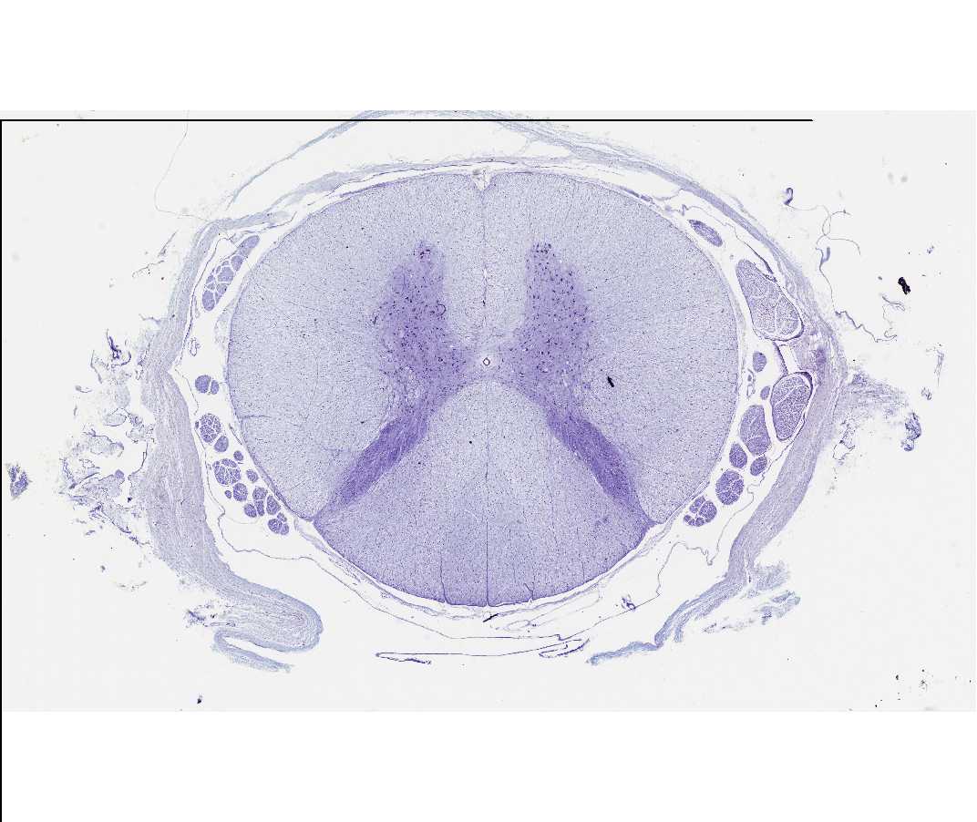
Identify the centrally located butterfly or H-shaped arrangement of the gray matter. Distinguish the white and gray matter, and the dorsal (posterior) and ventral (anterior) horns of the gray matter. Note: In this scanned image, ventral is at the top.
Under medium power, identify the cell bodies of the large motor neurons in the anterior horn of the gray matter. Identify the basophilic Nissl substance. Is there Nissl substance in the dendrites? In the axons? To what structures at the electron microscopic level do the Nissl bodies correspond?
Within the white matter, note the nuclei of glial cells (mostly oligodendroglia) and the cross sections of axons (unstained). The clear space surrounding each axon is occupied in life by the myelin sheath. Note the meninges surrounding the spinal cord. In some slides you will be able to identify the dorsal and ventral rootlets of the spinal nerves within the subarachnoid space.
#84 Spinal Cord, Monkey, Cross Section (Cajal's silver)
Open with WebViewer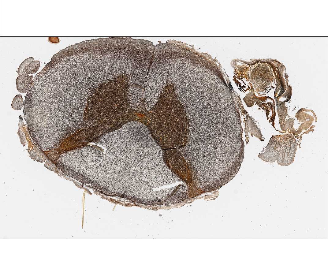
Identify the centrally located butterfly - shaped arrangement of the gray matter. Within the gray matter and locate the cell bodies (perikarya) of neurons, and the associated dendrites and axons. Surrounding the gray matter is the paler staining white matter and the supporting cells (oligodendroglia and others). In the gray matter, note the size and shape of the cell body (perikaryon) of the neurons, particularly those in the anterior (ventral) horn. With the Cajal technique, silver is precipitated on neurofilaments within neuronal cell bodies and their processes. In general, all of the cells and their processes are revealed by this technique. In the gray matter, note that most of the axons are oriented in the plane of the section. In contrast, most of the axons of the white matter are viewed in cross section, since the fibers are running to and from the brain and other segmental levels of the spinal cord. Note the surrounding meninges, and identify the central canal of the spinal cord.