SBPMD Histology Laboratory Manual
Urinary System: Kidney
The kidney is divided into lobes. One lobe consists of the conical medullary pyramid and the cortical substance that surrounds it like a cap. The guinea pig has a unilobar or unipyramidal kidney. The human kidney is composed of 12-13 pyramids.
#50 Kidney, Guinea Pig H&E
Open with WebViewer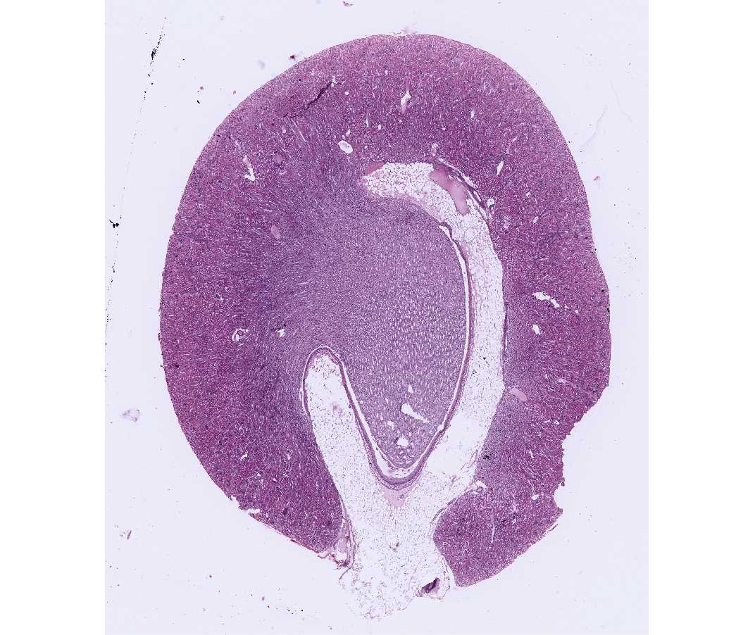
Identify the outer, brighter cortex with its glomeruli and the central, paler staining medulla. The base of the medullary pyramid lies below the cortex and the apex of the pyramid projects or empties into the renal pelvis. The hilus of the kidney is the site of entrance/exit of the renal artery, vein and ureter. Note the abundance of white fat in this region.
Examine the junction between cortex and medulla with the scanning objective. This junction is irregular. The cortex is subdivided into alternating regions: 1) the cortical labyrinth consists of glomeruli and convoluted tubules; and 2) the medullary rays consist primarily of radially directed straight segments of the loop of Henle and collecting tubules. A kidney lobule consists of a medullary ray and the portions of the adjacent cortical labyrinth. The medulla is further sub-divided into an outer zone adjacent to the cortex and an inner zone including the tip of the pyramid (which is called the papilla).
Under medium magnification and with the aid of the illustrations, examine the kidney in greater detail. Identify the different regions of the nephron, the structural and functional unit of the kidney. A nephron is composed of: 1) renal corpuscle, consisting of the vascular glomerulus and its capsule (Bowman's capsule); 2) proximal convoluted tubule; 3) loop of Henle, consisting of a thick descending segment, a thin U-shaped segment, and a thick ascending segment; and 4) distal convoluted tubule. The excretory portion of the kidney begins with the collecting tubules (which are in continuity with the distal convoluted tubules).
- Renal corpuscle. Within the renal corpuscle, identify the vascular capillary tuft or glomerulus. Search for a renal corpuscle in which you can identify the vascular pole. This is the site of the entrance and exit of the afferent and efferent arterioles, respectively. Identify the visceral and parietal layers of Bowman's capsule, and try to locate a renal corpuscle in which the parietal layer of Bowman's capsule is in continuity with a proximal convoluted tubule. This junction is the urinary pole of the renal corpuscle, and it lies opposite the site of the vascular pole.
- Proximal convoluted tubules. These surround the glomeruli, and they are the most abundant tubules of the cortical labyrinth. The cuboidal epithelium of the proximal tubules is strongly acidophilic, in contrast to the lightly stained distal tubules. Note the different appearance of the sectioned profiles of the proximal tubules, the result of different planes of section passing through a highly convoluted tubule.
- Search for a glomerulus in which the last portion of the thick ascending limb of the distal tubule is closely apposed to the vascular pole of the renal corpuscle. That portion of the distal tubule in intimate apposition with the afferent arteriole shows modifications: cells are more columnar and there is an increased concentration of nuclei. This modified area of the thick ascending tubule is called the macula densa. The macula densa, together with the modified muscle cells (juxtaglomerular or JG cells) of the afferent arteriole, form the juxta-glomerular apparatus. The JG cells are secretory; they release an enzyme called renin (not to be confused with rennin, the digestive enzyme) into the blood stream.
- Distal convoluted tubules. Leading from the macula densa, the nephron becomes the distal convoluted tubule. The cuboidal epithelium of the tubules is palely stained with H&E. The distal tubules are much less numerous than the proximal tubules in the cortical labyrinth.
Next, examine the medullary rays adjacent to the labyrinth and the medulla itself. Identify:
- the radially running thick descending segment of the loop of Henle (cytologically similar in appearance to the proximal convoluted tubules with which they are continuous in the ray)
- the thin segment of the loop of Henle (in most cases, the simple squamous epithelium of these tubules cannot be distinguished from that of a capillary in the inner zone of the medulla)
- the thick ascending segment of the loop of Henle (cytologically similar in appearance to the distal convoluted tubules with which they are continuous); returns to the glomerulus of origin of the nephron and forms the macula densa (see description above)
- collecting tubules in the ray and in the medulla. These latter tubules are pale-staining like the distal tubules, but differ from them in that their epithelium is more columnar, the apex of the epithelial cells tend to bulge into the tubule lumen, and the intercellular boundaries are readily evident as the cells do not form interdigitations.
Try to visualize the spatial relationships of an entire nephron as you examine the cortical labyrinth and rays, and anticipate which components you would expect to find in each region.
The renal medulla consists primarily of collecting tubules and larger collecting ducts, thin segments of the loop of Henle, and, to a varying extent, the thick ascending and descending segments of the loop of Henle. The largest collecting ducts that open on the area cribrosa of the papilla are the papillary ducts (of Bellini) (not present on all slides). A fortunate section may pass through the openings of some of the papillary ducts into the renal pelvis. Note the epithelial type lining the renal pelvis. The calyx itself is lined by transitional epithelium.
#103 Kidney, guinea pig, Periodic acid Schiff (PAS) reaction and hematoxylin
Open with WebViewer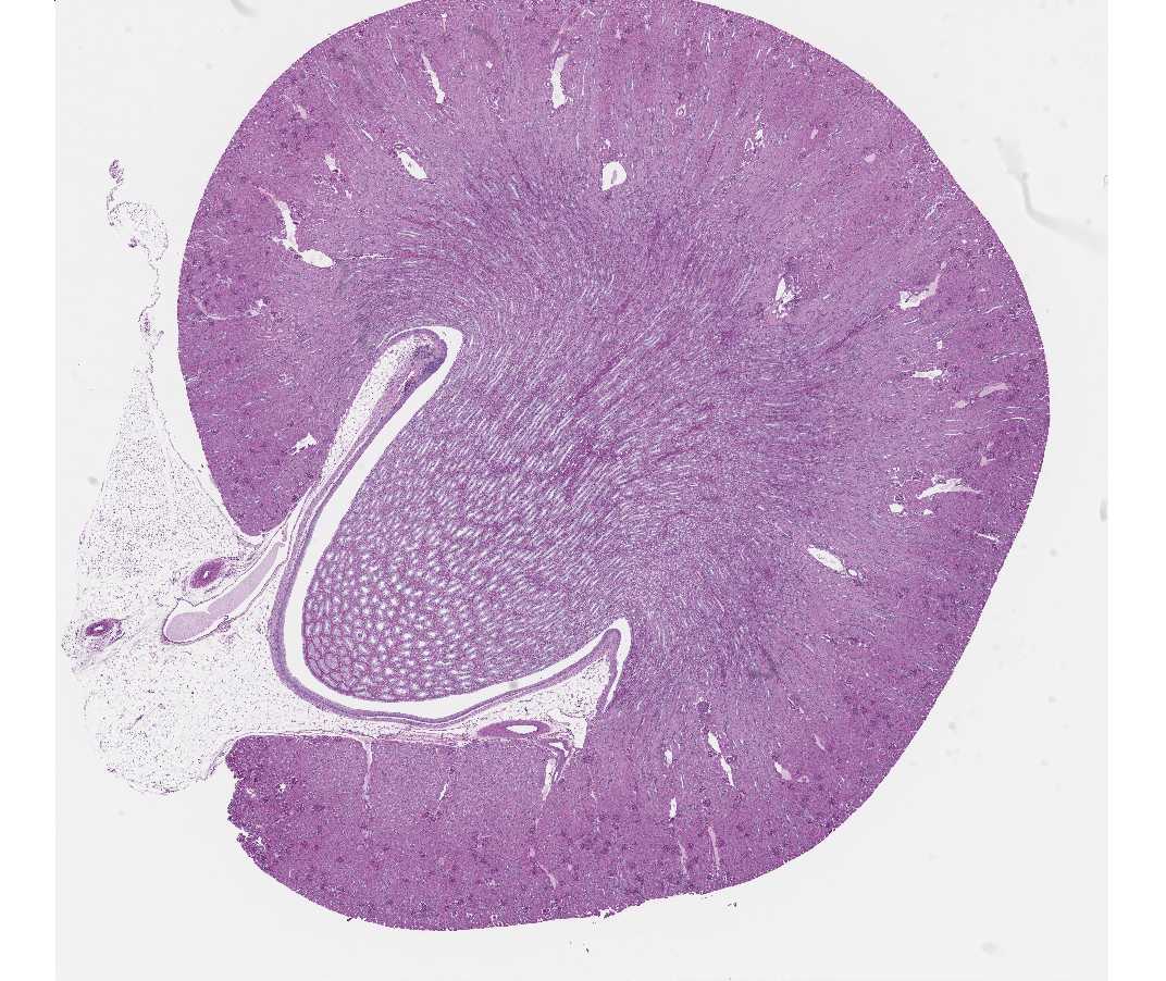
Orient yourself as for the previous slide, and then examine the cortical labyrinth and locate a region with several renal corpuscles. The staining differences of the general cytoplasm in the two types of convoluted tubules are not as distinct with this stain as with H&E, but the proximal convoluted tubules can be readily identified by the PAS-positive brush border (microvillus border) stained reddish or magenta at its luminal surface. Glomeruli, proximal and distal convoluted tubules are all sharply demarcated at their outer surfaces by PAS, since this is also an excellent stain for demonstrating basement membranes. Examine the tubules and glomeruli under higher magnification and identify all the components of the cortical labyrinth. Note especially regions in which the glomeruli have been section through the urinary and/or vascular pole. At the vascular pole, look for examples of the macula densa and the juxtaglomerular cells. Within the glomerulus, examine the parietal epithelium of Bowman's capsule and the visceral epithelium of the glomerulus. Be sure you understand the cells that form the visceral epithelium and the composition of the glomerular filter.
#51 Kidney, Guinea Pig, PAS & Hematoxylin (not scanned)
This is a thicker PAS-stained section than the section on slide #103. Identify structures as on #103.
Examine electron micrographs of the glomeruli, proximal and distal convoluted tubules in your textbook and in the lab, and correlate the PAS-positive structures evident with the light microscope with their ultrastructural counterparts. What is the functional significance of the occurrence of a brush border in the proximal tubule? Be sure you understand the significance of PAS staining.
#49 Kidney, Human, H&E.
Open with WebViewer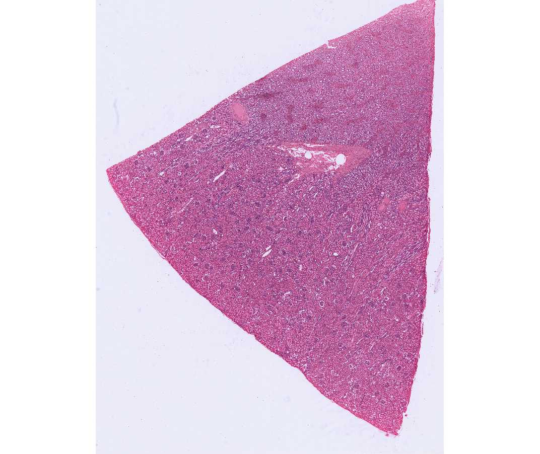
This wedge-shaped section represents only a small portion of the cortex of the human kidney. The human kidney is multilobar (multipyramidal), in contrast to the unilobar condition in the guinea pig. Examine the cortical labyrinth and rays as described previously.
Be certain that you understand the blood supply of the renal corpuscle, the convoluted tubules, and the loop of Henle, and the functional significance of these. Consult your textbook and its illustrations. What are the components of the arterial portal system of the kidney?
#1 Kidney. Rat. Regaud's iron hematoxylin, after dichromate fixation (not scanned)
Mitochondria are well demonstrated and can be seen clearly as black rods with the high-dry and oil immersion objectives. The filamentous mitochondria are oriented parallel to the lateral surfaces of the cell. Red blood cells and nucleoli are also stained. What is the significance of the differential distribution of mitochondria in the proximal versus the distal convoluted tubules?