SBPMD Histology Laboratory Manual
Gastrointestinal System I: Large Intestine
#39 Colon, Monkey, thin plastic section (H&E)
Open with WebViewer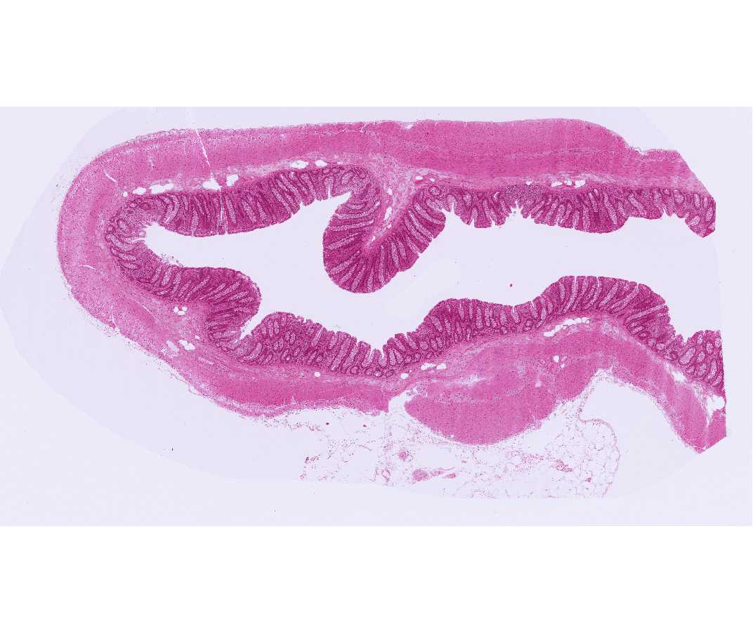
Note that the mucosa contains crypts and lacks villi. Also note the abundance of goblet cells in the lining of the crypts but not on the luminal surface; these features are diagnostic. This is a good slide in which to review the structure of arteries, veins and the peripheral autonomic plexus. Note the cells with eosinophilic granules (enteroendocrine cells) in the lining epithelium.
#48 Rectum and Anal Canal, Human
Open with WebViewer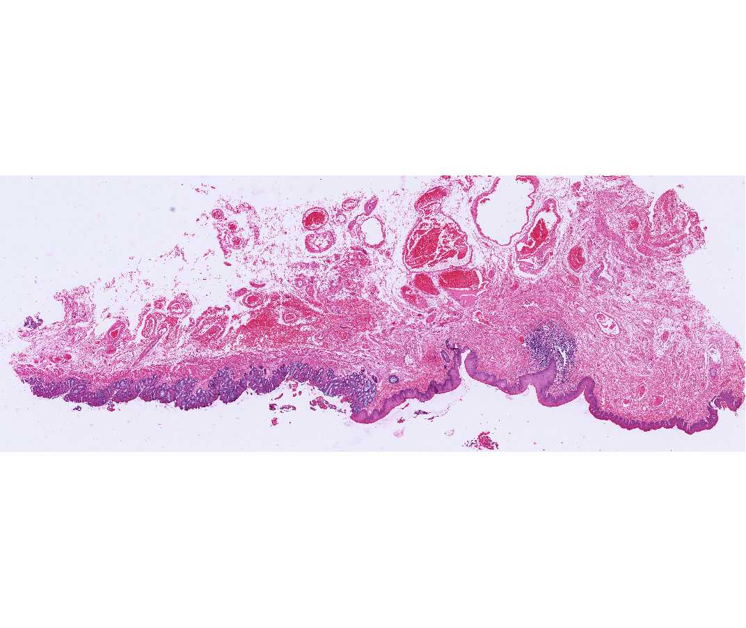
The rectum is histologically similar to the colon, and there is an abrupt transition between the rectal simple columnar epithelium and the stratified epithelium of the anal canal. The anal epithelium may appear stratified cuboidal at the junction with the rectum, but it assumes a typical stratified squamous appearance more distally. The intestinal glands end abruptly at the recto-anal junction. The inner circular layer of the muscularis externa is thickened considerably to form the internal anal sphincter. The submucosa of the anal canal is characterized by an extensive plexus of hemorrhoidal vessels. The abnormal dilation and varicosity of these vessels causes an inward bulging of the mucous membrane and a partial occlusion of the anal canal, resulting in internal hemorrhoids.
#106 Appendix, Human, H&E (scanned only, no slide in box)
Open with WebViewer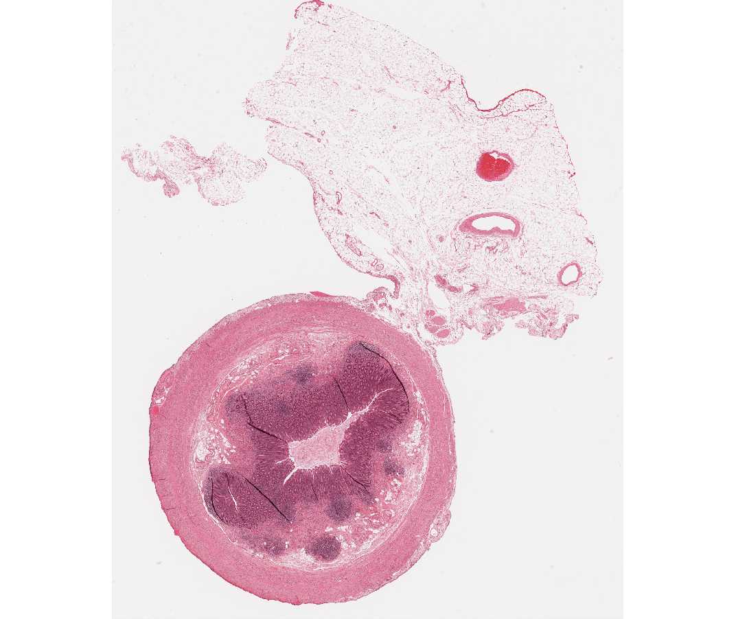
Examine the components of the wall of this portion of the gastrointestinal tract and compare them with the small and large intestine. In the mucosa of the appendix there is extensive GALT that extends into the submucosa. Both diffuse and nodular lymphatic tissue are present.