SBPMD Histology Laboratory Manual
Gastrointestinal System I
Examine the electron Micrographs so that you understand the ultrastructural equivalents of the structures you have seen under the microscope.
Parietal cell, stomach
Click to see enlarged view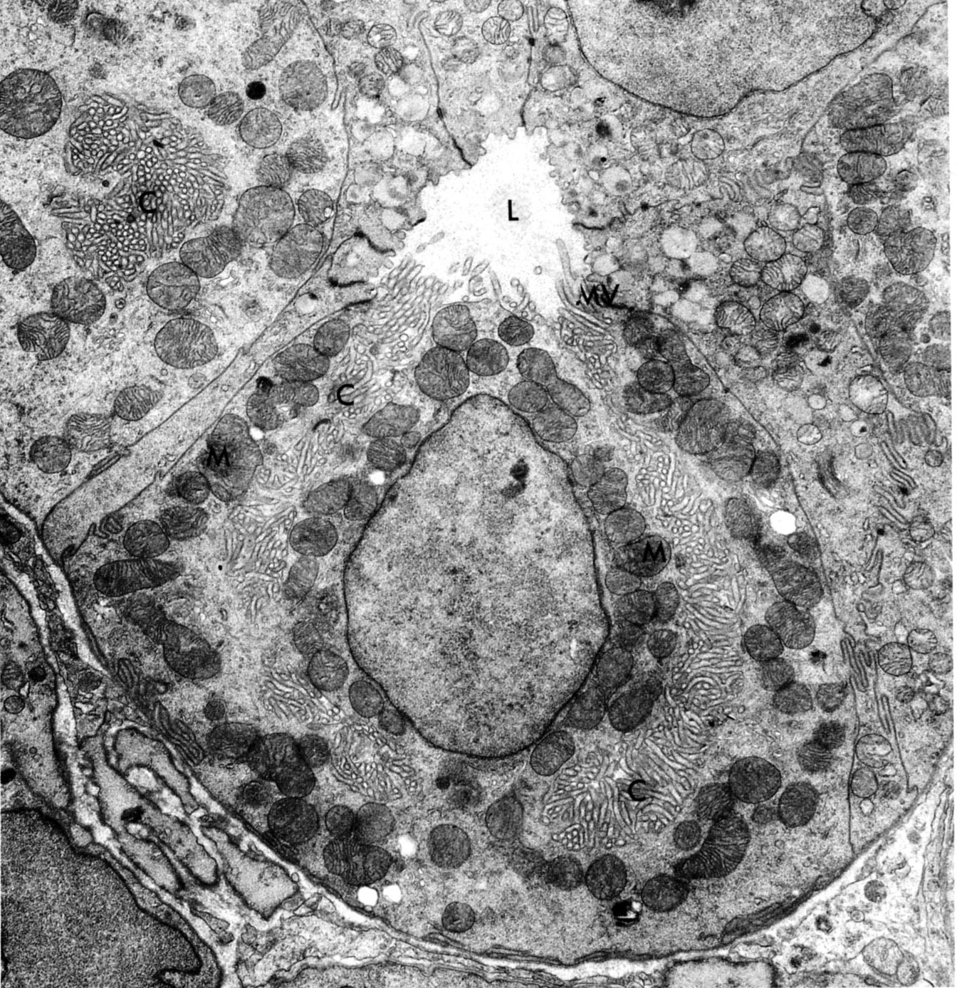
Parietal cell in gastric gland of bat whose stomach has been stimulated to produce hydrochloric acid. The secretory canaliculus (C) is a deep, troughlike invagination of the apical cell surface opening into the lumen (L). Its surface area is further increased by numerous microvilli (MV). Mitochondria are abundant.
Weiss, L. ed., Cell and Tissue Biology, 6th ed., Urban & Schwarzenberg, Baltimore, 1988, p. 654.
Chief cell in gastric gland of monkey
Click to see enlarged view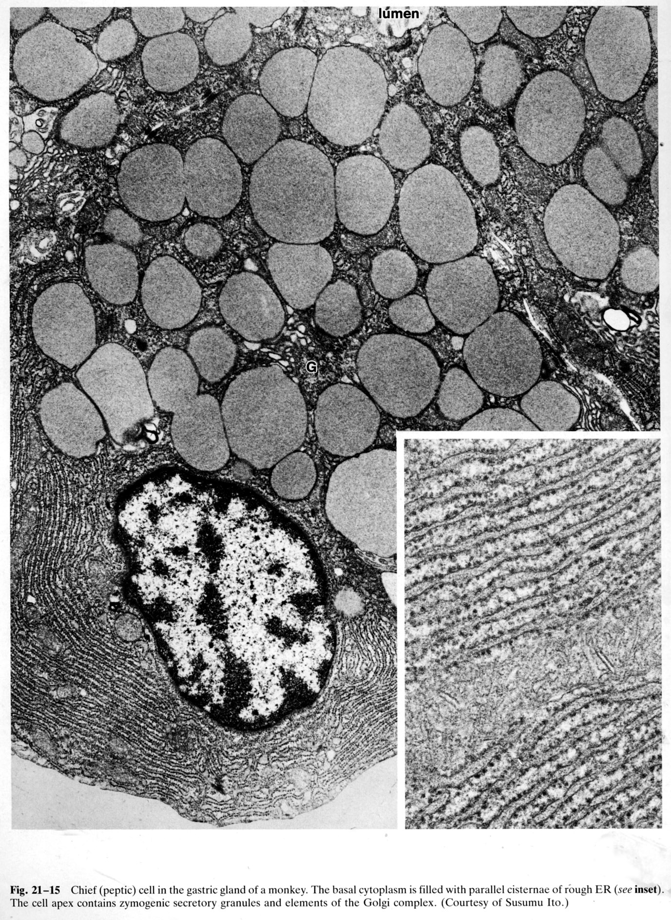
The basal cytoplasm is filled with parallel cisternae of RER (see inset). The apex of the cell contains zymogenic secretory granules and elements of the Golgi complex.
Weiss, L. ed., Cell and Tissue Biology, 6th ed., Urban & Schwarzenberg, Baltimore, 1988, p. 656.
Epithelial lining of small intestine
Click to see enlarged view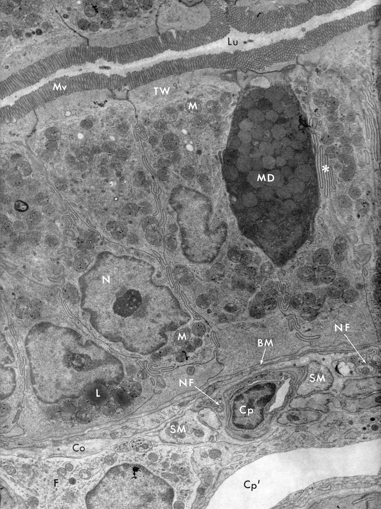
Simple columnar epithelium lining of the small intestine made up of absorptive cells (enterocytes) and a goblet cell containing mucous droplets (MD). (Neither the apical region of the goblet cell nor its nucleus is in the plane of section.) Note the microvillous border (Mv) on the luminal surface (Lu) of the enterocytes and the region of the terminal web (TW). The lateral cell borders are interdigitating (asterisk) and the cells rest on a basal lamina (BM). The lamina propria contains capillaries cut in cross section (Cp) and in longitudinal section (Cp), smooth muscle cells (SM), nerve fibers (NF), fibroblasts (F), collagen fibers (Co). Nucleus (N), lipid droplet (L), mitochondria (M). (duodenum of bat)
Porter, KR and Bonneville, MA, Fine Structure of Cells and Tissues, 4th ed., Lea & Febiger, Philadelphia, 1973, p. 28.
Apical region of intestinal epithelial cell
Click to see enlarged view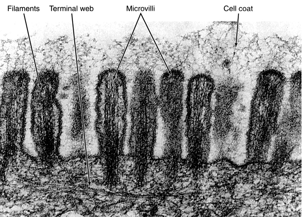
An extracellular coat (glycocalyx) is bound to the plasmalemma of the microvilli of enterocytes. The glycoproteins in this case include terminal digestive enzymes such as dipeptidases and disaccharidases. Actin filaments that constitute the core of the microvilli are clearly seen.
Junqueira, LC and Carneiro, J, Basic Histology 11th ed., McGraw-Hill, New York, 2005. P. 72.
Goblet cell
Click to see enlarged view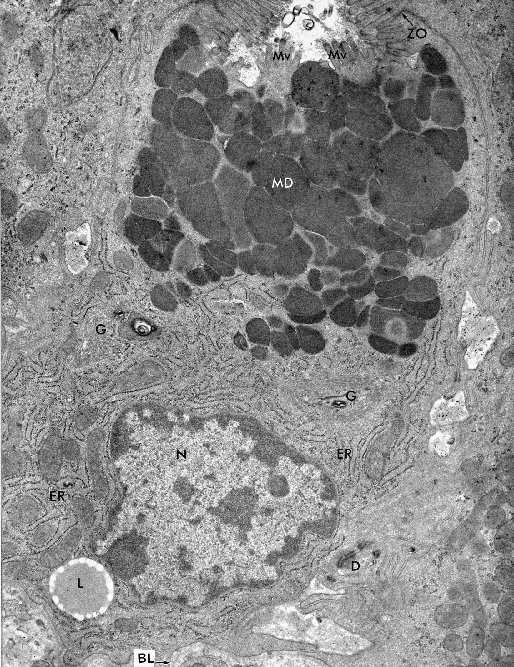
A goblet cell (small intestine, bat) with closely packed mucous droplets (MD). One droplet appears to be close to exiting the cell between two clusters of microvilli (Mv). The apical region of the cell is joined to its neighbor by a zonula occludens (ZO). The nucleus (N) is basal and there is abundant rough endoplasmic reticulum (ER). Elements of the Golgi can also be seen (G). Lipid inclusions (L) are common. These cells rest on the basal lamina (BL) and desmosomes (D) occur at the basolateral surface.
Porter, KR and Bonneville, MA, Fine Structure of Cells and Tissues, 4th ed., Lea & Febiger, Philadelphia, 1973, p.54.
Enteroendocrine cell of gut
Click to see enlarged view
Endocrine cell in gastric gland of a bat, situated between two chief cells (C ). Its basal surface, the site of hormone release, rests against basal lamina (bl). The cytoplasm contains small, dense granules (arrows). Product is released basally to be conveyed by the vasculature.
Weiss, L. ed., Cell and Tissue Biology, 6th ed., Urban & Schwarzenberg, Baltimore, 1988, p. 657.