SBPMD Histology Laboratory Manual
Gastrointestinal System I: Small Intestine
#35 Stomach and Duodenum, Human or Monkey
Open with WebViewer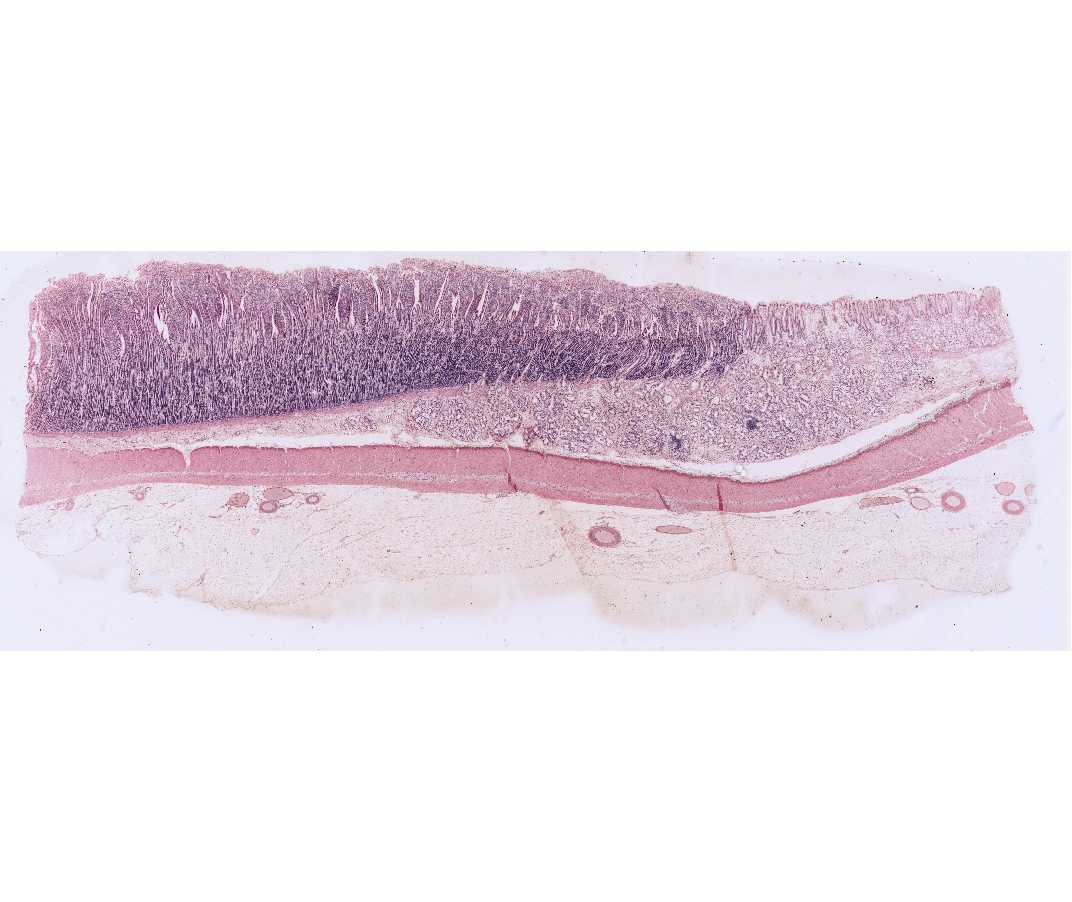
Locate the junction of the stomach and duodenum. Look for differences in the epithelial surface and, particularly on the monkey slide, note the thickening of the muscularis externa of the stomach as it becomes the pyloric sphincter. Using higher magnification, locate the junction by examining the mucosa, particularly the surface epithelium. The mucosal surface of the duodenum is thrown up into grossly visible plicae circulares (circular folds) by the elevation of submucosal folds. Although the gastric mucosa is characterized by surface pits and the intestinal mucosa is characterized by finger-like outpockets, or villi, this distinction is not always readily apparent on sections. One of the best ways to distinguish between the two organs is to examine the surface epithelium that lines the pits or villi. In the stomach the cells all have a basically uniform appearance, since they are all mucus-secreting cells. In the intestinal villi, however, most of the cells are absorptive cells, and interspersed between these are the characteristic mucous-secreting goblet cells. Goblet cells of the intestine will stand out when the slide is scanned under low power. In addition, a striate border can sometimes be seen on the free surface of the absorptive cells in well-preserved intestinal villi. To what ultrastructural feature does the striated border correspond? The duodenum is also characterized by the presence of mucus-secreting duodenal glands (of Brunner) in its submucosa. Unfortunately, in this slide, there has been considerable autolysis of the surface of the intestinal villi.
#36 Small intestine, duodenum, Human
Open with WebViewer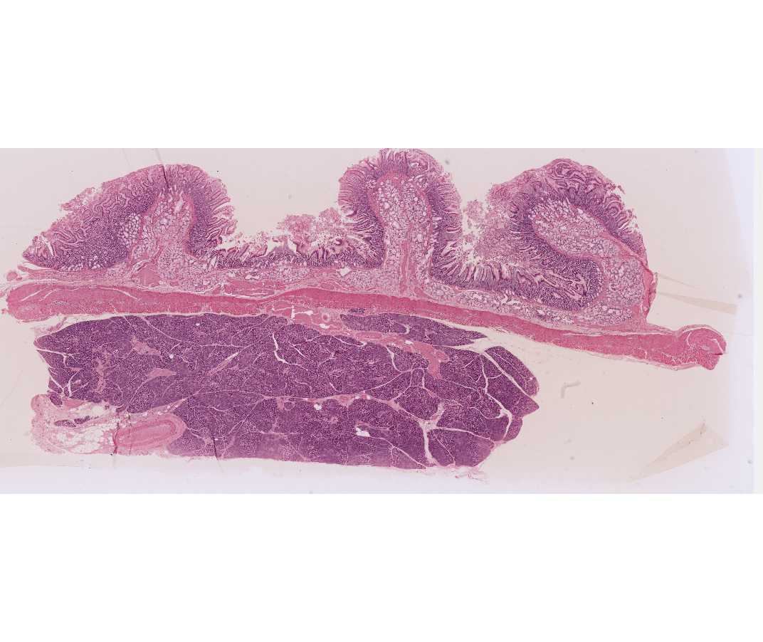
Identify the three components of the mucosa: epithelium, lamina propria and muscularis mucosae. Note the submucosal glands of Bruner. The two layers of the muscularis externa are present and outside these is the adventitia. Between the two layers of the muscularis externa, identify elements of the myenteric plexus. In most slides some pancreas can be seen beneath the muscularis externa. This will be studied in the next lab.
#37 and #117 Small Intestine, Human, Dog or Rabbit, H&E. (#37 not scanned)
Open with WebViewer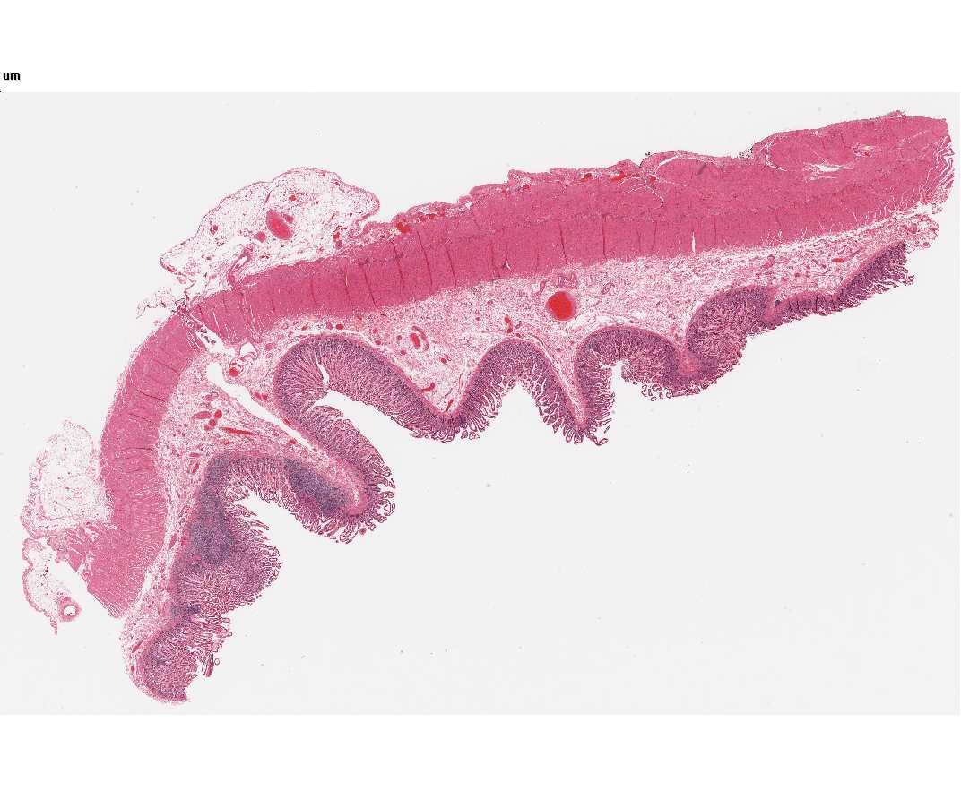
Identify the components of the wall of the small intestine. Note the presence of diffuse lymphatic tissue in the mucosa (GALT). (In the ileum there are accumulations of lymph nodules called Peyer’s patches). At the base of the intestinal crypts are Paneth cells with accumulations of large acidophilic granules in their apical cytoplasm, and strongly basophilic basal cytoplasm. The appearance of these cells is characteristic of enzyme-secreting cells. These cells secrete the anti-bacterial enzyme lysozyme.
#102 Small Intestine. Guinea Pig (Bodian/silver)
Open with WebViewer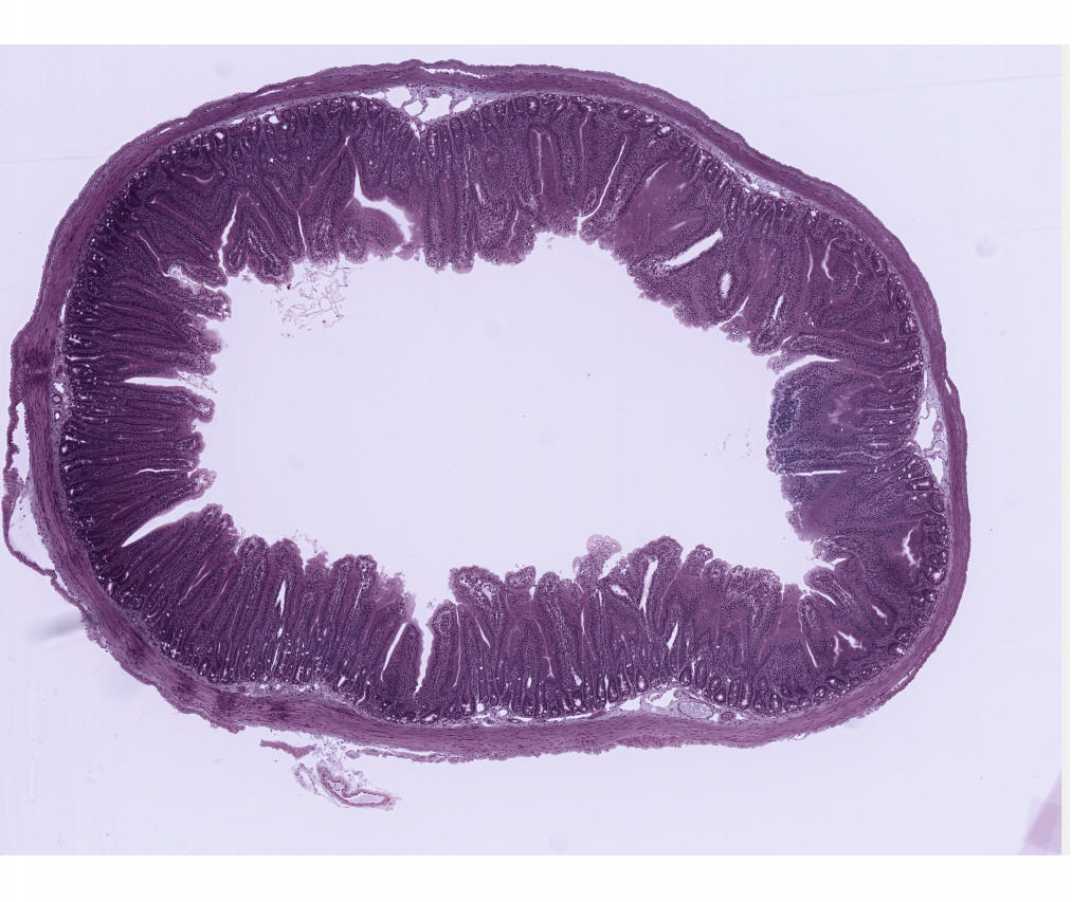
The striated border (microvilli) and terminal bar (junctional complex) are demonstrated particularly well on this slide. In addition, the Bodian silver stain demonstrates the enteroendocrine cells (use high magnification). What are the secretory products of these cells? What is their possible function? In what other regions of the digestive tract are they found? Note the basal lamina around smooth muscle cells in both longitudinal and cross-section. Myenteric neurons are also very nicely demonstrated in this slide.
#101a Small Intestine. Guinea Pig (PAS and hematoxylin)
Open with WebViewer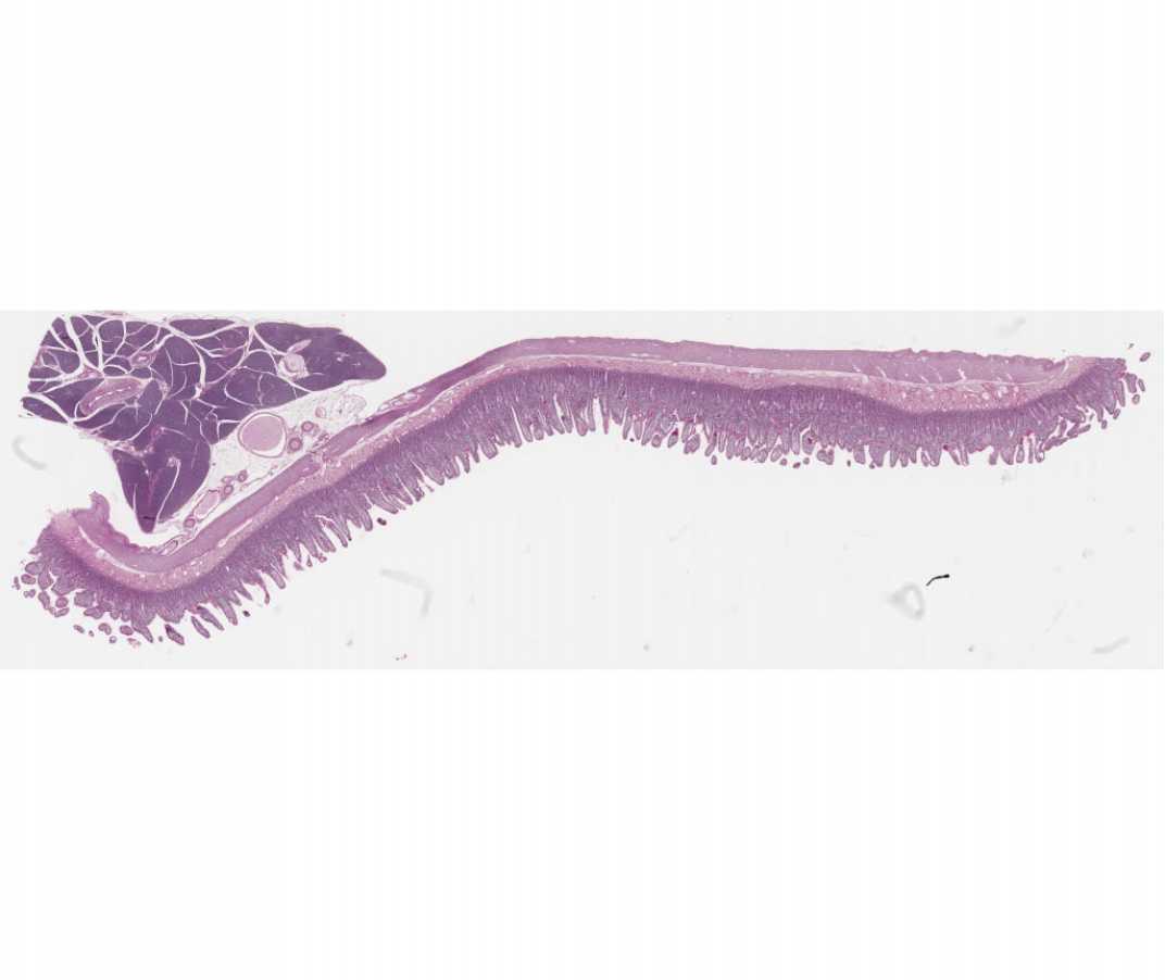
This slide is useful for demonstrating structures that contain glycosaminoglycans. The glycocalyx that covers the microvillus border of villus absorptive cells and intensely stained goblet cells can be seen. Within the crypts of Lieberkuhn, which contain undifferentiated cells, the glycocalyx is not present. Note also the staining in the loose connective tissue of the lamina propria and the intense staining of goblet cells in the epithelium.
#38 Small Intestine, Monkey, Fat Absorption
Demonstration Slide
This slide is a beautiful presentation of the mechanism of lipid absorption in the small intestine. The ultrastructural mechanisms of lipid absorption will be discussed in lecture, but the general pathways of lipid uptake are evident on your slide. Osmium is dissolved within the lipid droplets and renders them insoluble and visible. Consider the occurrence of lipid droplets in the following sequence: (1) in the intestinal lumen; (2) adhering to the surface of the striate (microvillus) border or located as a dark line at the base of the striations; (3) in large aggregations of droplets enclosed in SER within the apical cytoplasm just beneath the striate border; (4) in droplets within the intercellular spaces between the lateral margins of the epithelial cells, particularly toward the basal half of the cells; (5) within the interstitial spaces of the loose connective tissue of the lamina propria that forms the core of the villi; and (6) within the lumen of the distended central lacteals.