SBPMD Histology Laboratory Manual
Gastrointestinal System I: Stomach
The stomach extends from the esophagus to the duodenum; it is divisible into the cardiac, fundus, body, and pyloric regions. The distensible stomach is involved in both the mechanical and chemical breakdown of food, and also serves as a temporary reservoir. Its epithelium is specialized for secretion and is of the simple columnar type. The gastric mucosa contains gastric pits (foveolae); these are surface invaginations that also serve as the ducts of the underlying intrinsic gastric glands. Three basic cell types contribute to the secretion of gastric juice, and each has a characteristic appearance under the light and electron microscope. All of these cell types can be seen in the fundus and body of the stomach.
- Mucus-secreting cells. These cells form the surface epithelium and extend inward to line the gastric pits. Nuclei are basal, and the supranuclear cytoplasm containing mucinogen granules appears clear or vacuolated with H & E stain. Mucous neck cells occur in the junctional region of the gastric pits and glands, and it is in this region that cell proliferation for the renewal of the epithelium occurs.
- Parietal cells. These pyramidal or spherical cells appear wedged in between other cells of the gastric glands. They are characterized by their finely granular acidophilic cytoplasm due to an abundance of mitochondria, and by their central, spherical nucleus. They contain an extensive intracellular canalicular system that communicates with the lumen of the gland (visible at the EM level). These cells are involved in the elaboration of hydrochloric acid and intrinsic factor.
- Chief (zymogen) cells. As their name implies, these cells are involved in the secretion of enzymes, particularly the proteolytic enzyme pepsinogen (pepsin in the active state). As is characteristic of cells involved in protein synthesis and secretion, these cells contain basophilic cytoplasm, particularly at their base due to the extensive development of rough endoplasmic reticulum. Although at the LM level the supranuclear cytoplasm generally appears clear or vacuolated, electron micrographs reveal an accumulation of membrane-bound secretory granules in this region.
#34 Stomach, Body, Monkey, H&E
Open with WebViewer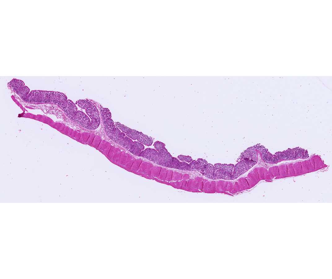
Identify the mucosa, submucosa, and muscularis externa. Locate the following elements of the mucosa: the luminal surface mucous secreting cells, the gastric pits and the cells lining them. Parietal cells are particularly prominent and chief cells and mucous neck cells are present. Note the loose, cellular areolar connective tissue surrounding the gastric pits, the muscularis mucosae, which forms a boundary between the mucosa and submucosa, and the blood vessels in the submucosa. Find nerve bundles and the ganglion cells of the submucosal plexus (Meissner's plexus), but they are not obvious in all of them. Examine the muscularis externa and notice that the smooth muscle is oriented in several different planes. A serosa covers the external surface of the gland in this section. Try to find the myenteric plexus (Auerbach's plexus) between the external and adjacent inner layers of smooth muscle.