SBPMD Histology Laboratory Manual
The Ear
Learning objectives:
- Understand the bony and membranous labyrinths.
- In the vestibule be able to recognize and describe the function of the sensory end organs.
- Be able to recognize and describe the function of the components of the cochlea including the organ of Corti.
#120 Ear, cochlea and vestibule
Open with WebViewer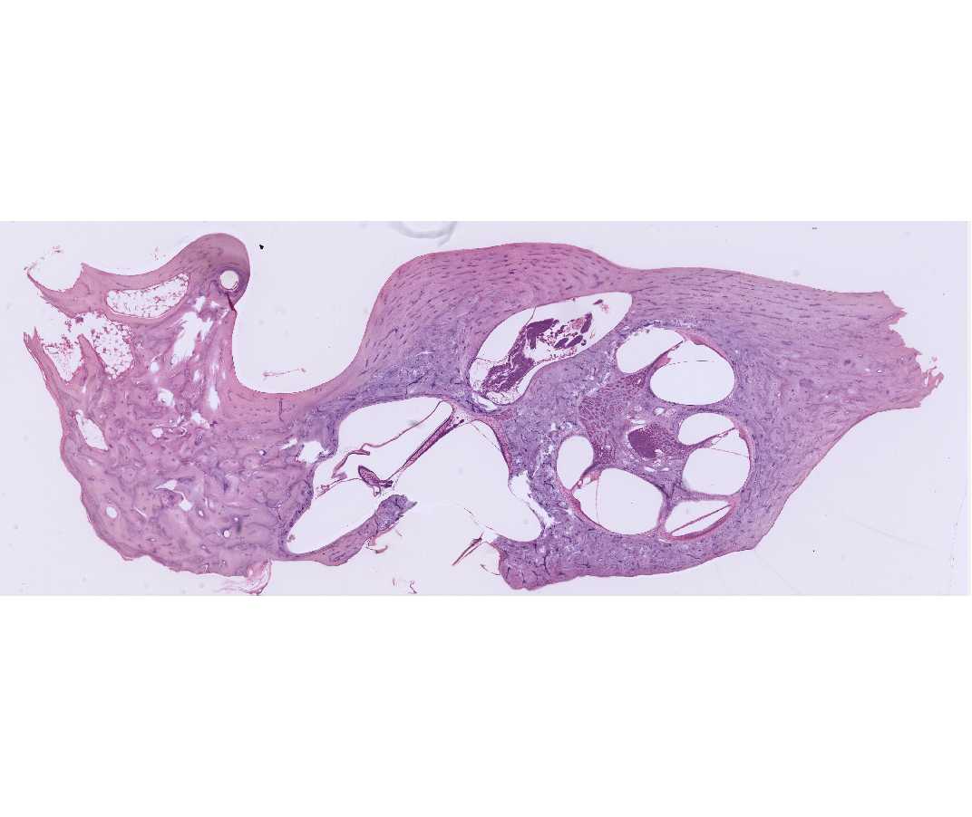
The ear presents some difficulties not encountered in previous slides. First, the sections are celloidin rather than paraffin and so are rather thick (about 20 mm), and second, the cochlea is a coiled structure and so is always sectioned repeatedly and often oddly. To get the most from the slide proceed as follows:
The bony labyrinth is a system of communicating cavities within the temporal bone, which are divided into three major regions: the semicircular canals (superior, horizontal and posterior), the vestibule and the cochlea. Suspended within the osseous labyrinth is the membranous labyrinth, which contains endolymph. Its wall is separated from the wall of the osseous labyrinth by a space containing perilymph. The membranous labyrinth consists of three semicircular canals, two small sacs, the utricle and saccule, and the scala media of the cochlear duct, all of which are in communication with each other.
Find the cochlea and the vestibule.
- Under low power (4X) locate:
- Temporal bone
- Fibers of cranial nerve VIII (cochlear or acoustic nerve, both of which contain both afferent and efferent fibers)
- Vestibule
- Cochlea
- Modiolus
- Spiral ganglia
- Under low power (10X) locate:
The turns of the cochlear duct
Within the cochlea- Modiolus - the central core of the cochlea, formed of spongy bone
- Spiral ganglion - cell bodies of sensory neurons (bipolar neurons) of the cochlear nerve (a division of cranial nerve VIII) found within the modiolus. Depending upon the plane of section, there will be portions of the spiral ganglion at each turn of the cochlea
- Osseous spiral lamina - a spiraling shelf of bone projecting from the modiolus
- Three chambers of cochlear duct:
- Scala media - cochlear duct (membranous labyrinth) - the triangular- shaped space whose acute angle is attached to the osseous spiral lamina
- Scala vestibuli - begins at oval window, is confluent with scala tympani at the helicotrema
- Scala tympani - begins at the helicotrema, ends at the round window
- Membranes separating the scalae
- Basilar membrane - supports the organ of Corti and divides the scala
media from the scala tympani - Vestibular membrane (Reissner's Membrane) divides the scala media from the scala vestibuli, arises medially.
- Basilar membrane - supports the organ of Corti and divides the scala
- Spiral ligament - a thickening of the periosteum on the outer wall of the scala media
- Under high power within the cochlea identify:
- Outer spiral ligament - periosteum of outer wall of cochlear duct
- Spiral prominence - thickened region of outer spiral ligament adjacent to stria vascularis
- Stria vascularis - portion of spiral ligament lined by vascularized stratified epithelium, secretes endolymph
- Bipolar ganglion cells of spiral ganglion and their processes
- The organ of Corti.
- Limbus spiralis - periosteal connective tissue at the inner angle of the scala media from which the vestibular lip protrudes
- Vestibular lip - pointed tip of the limbus spiralis from which the tectorial membrane arises
- Tectorial membrane - secretory product of cells of vestibular lip (proteoglycans and fine fibers), overlying organ of Corti
- Inner border cells - lining of internal spiral tunnel (inner spiral sulcus) from vestibular lip to inner hair cells
- Inner hair cells - single row of hair cells near internal spiral lamina
- Phalangeal cells (inner and outer) - support inner and outer hair cells
- Outer hair cells - external hair cells, arranged in three rows
- Inner and outer pillar cells - form borders of inner tunnel
- Inner tunnel - space formed by inner and outer pillar cells across which fibers of the cochlear nerve pass
- Supporting cells external to outer phalangeal cells (cells of Hensen, Claudius and Boettcher)
- Outer tunnel - space between outer phalangeal cells and cells of Hensen
- Habenula perforata – this is the region in the osseous spiral lamina where nerves enter and exit the organ of Cort
- Within the vestibule identify:
- Utricle - the portion of the membranous labyrinth into which the semicircular canals open
- Saccule - the portion of the membranous labyrinth which is continuous with the cochlear duct
- Identify the following specialized structures containing receptors for movement within the utricle or saccule:
Maculae - sensory organs of utricle and saccule. Macula of saccule is attached to periosteum while that of the utricle is on wall of membranous labyrinth.- Hair cells (Note that the two types, I and II, cannot be distinguished.)
- Otolithic membrane
- Phalangeal cells
- At the ampullae of the semicircular canals (adjacent to the utricle):
Crista ampullaris - sense organ found at each of the openings of the semicircular canals to the utricle. Not all slides have a crista ampullaris. Identify:- Hair cells (Note that the two types, I and II, cannot be distinguished.)
- Cupula (missing in many slides)
- Phalangeal cells