SBPMD Histology Laboratory Manual
Male Reproductive System: Micrograph
Examine the electron Micrographs so that you understand the ultrastructural equivalents of the structures you have seen under the microscope.
Spermiogenesis (sperm maturation), Golgi phase | |
| Click to see enlarged view | |
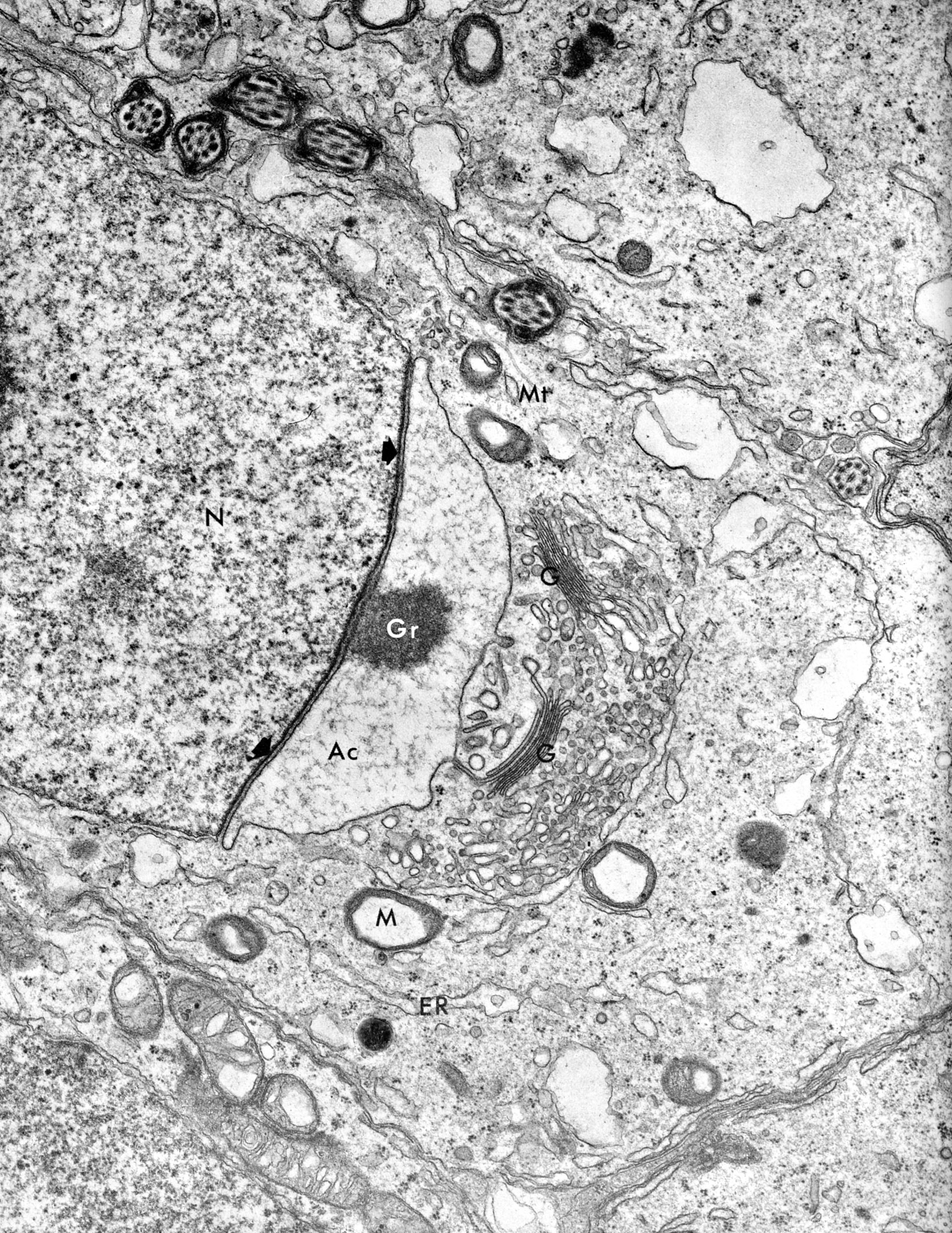 |
In the Golgi phase of spermiogenesis multiple Golgi complexes (G) accumulate. Proacrosomal granules (Gr) of these coalesce to form the acrosomal vesicle (Ac). This then spreads over the anterior half of the nucleus (N), forming the acrosomal cap. Mitochondria (M), microtubules (Mt), endoplasmic reticulum (ER).(testis mouse) |
| |
Spermiogenesis, spermatids | |
| Click to see enlarged view | |
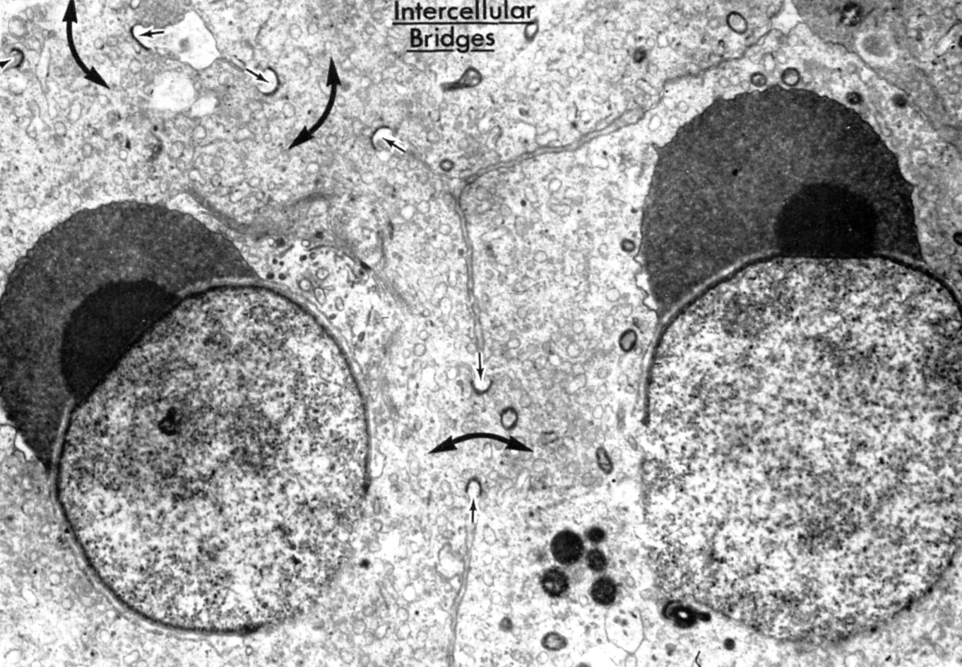 |
Portions of two spermatids (guinea pig) showing the intercellular bridges by which they are joined to each other and to two other spermatids of the same cluster. The small arrows indicate the local thickening of the cell membrane encircling the bridges. The large arrows passing through the bridges indicate the sites of continuity of the cytoplasm from cell to cell. Acrosome is forming adjacent to nucleus. |
| |
Spermiogenesis, spermatid (almost mature) | |
| Click to see enlarged view | |
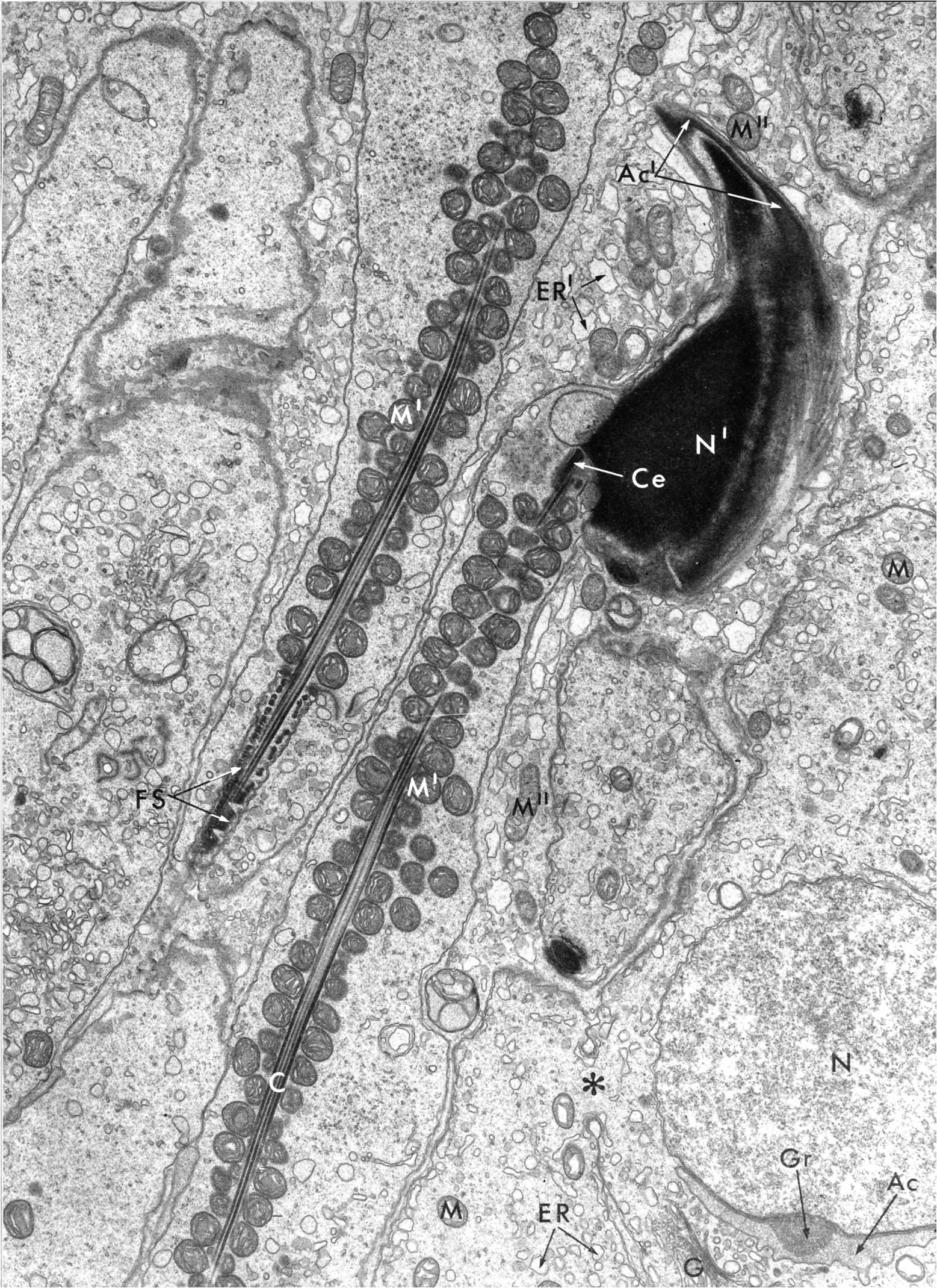 |
Nearly mature sperm with head (hook-shaped in this case, mouse) contains the nucleus (N), topped by the acrosome (Ac). Two centrioles are involved in the formation of the neck region. One of these (Ce) initiates the development of the long bundle of filaments (C ) that is the core of the middle piece and tail. The core has an internal structure similar to an cilium. The middle piece of the flagellum is wrapped by a sheath of mitochondria (M). The tail proper is a fibrous sheath (FS) wound around the axial filamenture. The asterisk indicates a cellular bridge between spermatids. Mitochondria (M), endoplasmic reticulum (ER). |
| |