SBPMD Histology Laboratory Manual
Seminal Vesicle
#60 Seminal Vesicle, Human, Age 34, H&E
Open with WebViewer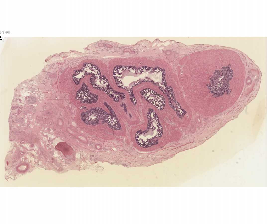
The highly folded lumen of the seminal vesicle appears as separate cavities when examined with the naked eye. With the microscope note that there are no discrete secretory alveoli in the seminal vesicle; instead, the entire lining membrane of the saccular gland is thrown into a series of complex, high, thin folds. The lining epithelium is generally simple columnar or pseudostratified, and basal cells are frequently seen, as in the epididymis and ductus deferens. The lamina propria contains connective tissue and smooth muscle cells. The seminal vesicle is embryologically derived from the ductus deferens, and like the latter, it has a prominent muscularis. This provides for the expulsion of seminal vesicle fluid during ejaculation. The acidophilic secretory material in the lumen of the gland is rich in fructose, and this apparently serves as an energy source for spermatozoa following ejaculation. Contrary to the implications of its name, the seminal vesicle is not a site of spermatozoa storage.