
 X. Edward Guo, Ph.D.


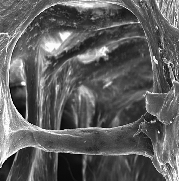
Characterization of in vivo trabecular bone adaptation by 3-D microimaging technique, quantification of in vivo histochemical consequences and their dependence on mechanical loading, and correlation of morphological and histochemical consequences to the local tissue strain variations using a microstructural model. This project utilizes an in vivo rat model for characterization of trabecular bone adaptation to mechanical loading. The aim is to establish a physical law that relates mechanical loading to biological adaptation of bone tissue.
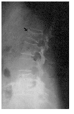
Failure patterns in vertebral bodies: Quantification of occurrence, location of compressive failure in vertebral bodies using mechanical testing, optical local strain measurements and microstructural modeling techniques. The aim of this project is to understand failure mechanisms at a whole bone level and correlating whole bone properties to microstructural features (individual trabeculae) using microstructural modeling techniques.
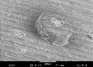
(1) Determination of interfacial debonding strength of cement lines using an osteon pushout test. The property of cement line has been hypothesized to play an important role in strength of cortical bone, and may also be a crucial factor for understanding lammelar structures in both cortical and trabecular bone tissues. The lamellar properties of bone tissue are crucial in determining mechanical properties at sub-microstructural level.
(2) Fracture Mechanics of Osteonal Cortical Bone: Application of fiber-matrix composite fracture mechanics methods to predict strength, fracture process in osteonal cortical bone. It has been long hypothesized that cortical bone behaves like a fiber-matrix composite material without any verification. The purpose of this study is to verify applicability of current fracture mechanics techniques for fiber-matrix composites to cortical bone, to quantify contributions of various microstructural components to fracture properties of cortical bone.
 BACK TO MAIN
 FORWARD TO PUBLICATIONS