 |
Jason J. Psillakis, DDS, MSa [MEDLINE LOOKUP]Sections
Mona E. McAlarney, DEngScb [MEDLINE LOOKUP]
Robert F. Wright, DDSc [MEDLINE LOOKUP]
Javier Urquiola, DDSd [MEDLINE LOOKUP]
School of Dental and Oral Surgery, Columbia University, New York, N.Y.
-
Abstract
-
Material and Methods
-
Results
-
Discussion
-
Conclusions
-
References
-
Publishing and Reprint Information
| Abstract | TOP |
Statement of problem. Casting relief is required for proper seating of castings to allow for luting agent thickness. The application of die spacer to the die is the most common method of obtaining casting relief. Die spacer film thicknesses that are outside the ideal range of 25 to 40 µm can cause clinical problems. Thickness can be affected by the separation of die spacer constituents, which may not be reconstituted by mixing, in the bottle and by the evaporation of volatile components while the bottle is open.
Purpose. The purpose of this study was to determine the effects of component evaporation and die spacer mixing technique on applied die spacer thickness.
Material and methods. Bottles of Gold Tru-fit die spacer were left open for 0, 1, 4, 8, and 24 hours at 22°C. Spacer solutions were shaken either by hand per the manufacturer's directions or on a dental vibrator for 1 minute. One even brush stroke of spacer was applied to clean glass slides. Three die spacer films were made for each combination of time and mixing technique. Eighteen thickness measurements per sample at various sites were recorded with profilometer tracings. Statistical differences were determined with a 2-way ANOVA.
Results. Handshaking provided greater die spacer thickness, which increased with the time that the bottle was open. Vibration provided lower thickness with no statistical increase with time.
Conclusion. Insufficient agitation caused lower film thickness. Excessive evaporation caused higher film thickness. (J Prosthet Dent 2001;85:82-7.)
| CLINICAL IMPLICATIONS The results of this preliminary study suggest that insufficient agitation during die spacer mixing may result in insufficient casting relief for proper seating of castings. Evaporation may have the opposite effect: bottles open for 4 or more hours are subject to the evaporation of organic liquid and may at least double the applied die spacer thickness, leading to excessive relief. |
The proper fit and cementation of a cast restoration onto a prepared tooth is crucial to both short- and long-term function. Casting fit is a function of the many variables related to changes in size and shape introduced by the materials used in the construction of a restoration; these variables can stem from both the intrinsic properties of the materials and the clinical technique. Even if all variables were controlled carefully to ensure a perfect fit, the restoration still could not be seated properly because of insufficient space for the luting agent.
Proper complete seating of full veneer restorations, without an excessive luting agent film thickness, is often difficult to achieve because of cement hydraulic pressure.1-7 Incomplete seating may result in occlusal interference after cementation, loss of proximal contact, reduced crown retention, discrepancy of marginal fit, secondary caries, plaque accumulation, periodontal problems, rapid dissolution of luting agents, and/or hypersensitivity.3-6,8-11
Internal relief, or venting of the casting, can overcome the effects of luting agent hydraulic pressure.2-9,12-19 The use of venting requires drilling holes in the casting. The casting is cemented in place and the vent holes filled. Although venting is an effective technique to improve the seating of the restoration, it has several disadvantages: An extra visit is necessary to fill the vent hole; the materials used to fill the vent hole may cause microleakage and/or discoloration; and, in the case of ceramometal restorations, occlusal venting may weaken the strength of the porcelain.
Internal relief avoids the limitations of venting. Internal relief provides a space between the casting and the abutment to accommodate the luting agent and the marginal expression of excess cement.20 Methods of obtaining internal relief include grinding the inside of the casting, internally carving the wax pattern, etching with aqua regia, electrochemical milling, cutting the internal channel, an d applying die spacer to the abutment.
The most popular method to achieve adequate internal relief is die spacing.3,8,9,16 Die spacer is a solution that is painted on the die before the fabrication of the pattern. The presence of the spacer provides space for the luting agent. The use of die spacer reduces elevation of the casting restorations,5,7,12,13,15,18,19,21-23 decreases seating time,7 improves outflow of excess cement,20 and lowers seating forces.7 Conflicting evidence exists on the effect of die spacer presence on clinical crown retention.7 Grinding, internal carving of the wax pattern, etching with aqua regia, and electrochemical milling are no longer recommended methods because they are inaccurate and inconsistent, and it is impossible to achieve uniform space for a cementing medium.3
The reported ideal die spacer thickness ranges from 25 to 40 µm.4,11,12,17,22-25 Die spacer thickness should be large enough to allow proper seating of the casting but not so large as to cause excessive cement thickness.26 For optimal results, die spacer thickness should be uniform.
In general, die spacers consist of metal-oxide powders and adhesives dispersed in an organic liquid such as a ketone. All components must be properly dispersed and the die spacer composition held constant for optimal clinical effectiveness. With time, particles tend to settle to the bottom of a standing bottle. If the bottle is open, the organic matrix liquid tends to evaporate because of its high vapor pressure, thereby changing the die spacer composition. Mixing technique and the overall time that a bottle is open during clinical use can affect die spacer thickness and therefore clinical success.
Manufacturers recommend shaking the die spacer bottle vigorously before application to adequately disperse the components. Replacing the cap on the die spacer bottle immediately after use and discarding well-used bottles are also clinically prudent.
To ensure a consistent film thickness during studies, researchers not only vigorously shake the bottle but also immediately cap the bottle after application and/or frequently replace the die spacer bottle. These optimal conditions of mixing and prevention of evaporation do not occur clinically, where bottles may stand open for some time and shaking is often less than vigorous.
The purpose of this preliminary study was to examine the effect of mixing intensity and evaporation of die spacer components on die spacer thickness. To this end, die spacer film thickness was measured after allowing die spacer components to evaporate by leaving the bottle open at room temperature for various times up to 24 hours and then mixing, either by hand (shaking) or by the vibration of the bottle.
| Material and methods | TOP |
A Gold Tru-fit spacer (George Taub Products, Jersey City, N.J.) bottle was left open for 0, 1, 4, 8, and 24 hours at 22°C. After each time interval, the bottles were closed (only for the mixing process) and mixed either by hand (shaking) according to the manufacturer's recommendations or by placing the bottle on a dental vibrator for 1 minute to disperse the material. After mixing, die spacer suspension was applied to clean glass slides with a single, even brush stroke. A new brush was used for each application. Samples were dried in air at 22°C. Three samples of die spacer suspension on glass slides were made for each combination of mixing technique and evaporation time variables.
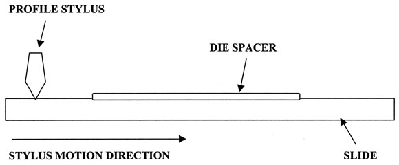
Die coating thickness was determined using a Tencor Alpha-Step 200 profilometer (KLA-Tencor Instruments, San Jose, Calif.) with a diamond stylus tip 12.5 µm in radius (Fig. 1).
|
Fig. 1. Schematic drawing of side view of slide coated with die spacer in profilometer. Profilometer stylus travels across slide from uncoated area to region coated with die spacer. When in contact with coating, stylus rises and records its elevation change as coating thickness. |
|
|
|
A profilometer measures film thickness by tracing a stylus from an uncoated region to a coated region. When the stylus comes in contact with the coating, it rises with the elevation of the film thickness. The change in stylus elevation due to the film is recorded.
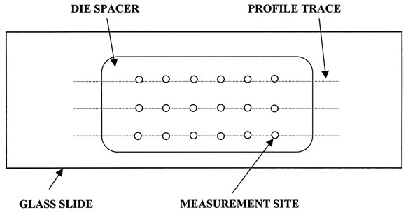
Three profile tracings were made for each sample (Fig. 2).
|
Fig. 2. Schematic drawing of top view of slide coated with die spacer. Dotted lines represent path of stylus across coating during profile trace. Open circles represent locations where film thickness measurements were made. |
)
|
|
|
For each tracing, 6 measurements of film thickness were made. These measurements were taken 250, 500, 750, 1000, 1250, and 1500 µm from the edge of the die spacer coating. Eighteen measurements (3 tracings × 6 measurements per tracing) were obtained for each sample. These 18 measurements were averaged to obtain 1 film thickness per sample.
A 2-way analysis of variance (ANOVA) was performed to determine the possible significance of any differences in film thickness with mixing technique or open bottle time (Sigma Stat, SPSS Inc, Chicago, Ill.). If a statistical difference was noted within a group, the Student-Newman-Keuls test with a set P .05 was used as a multiple comparison procedure to isolate any differences.
A composite plot of averaged die space film thickness values for all variables with standard deviation bars was constructed (Sigma Plot, SPSS Inc).
| Results | TOP |
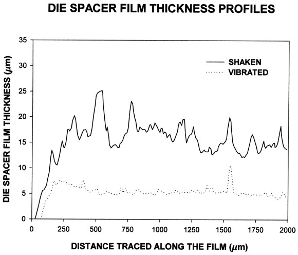
Typical thickness profiles for both handshaken and vibrated films are provided in Figure 3.
|
Fig. 3. Typical film thickness profiles recorded by profilometer during single trace along die spacer coating. Profiles are for handshaken and vibrated mixing for die spacer bottles open for 24 hours. |
|
|
|
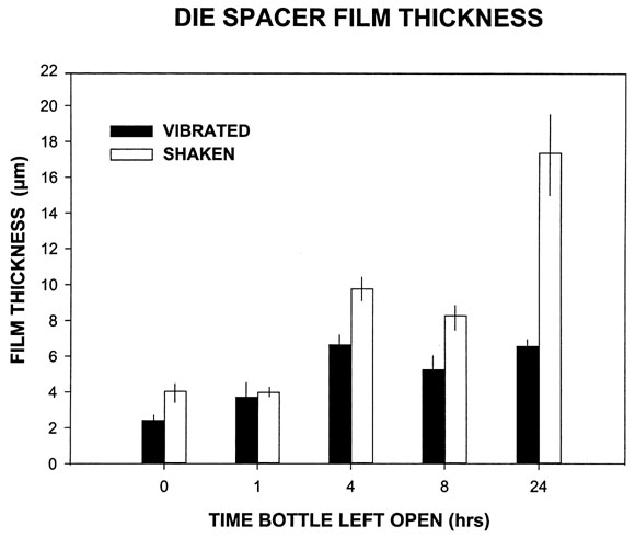
Mean values for each sample were obtained by averaging the thickness both over the 6 points along a single profile trace and over the 3 tracings per sample (Fig. 2![]() ). A plot of the effects of mixing method and open bottle time on die space film thickness was made (Fig. 4).
). A plot of the effects of mixing method and open bottle time on die space film thickness was made (Fig. 4).
|
Fig. 4. Plot of film thickness versus mixing mrethods and time die spacer bottle was left open. Bars represent averaged values; vertical lines on bars represent standard deviations. |
|
|
|
For mixing by hand (shaking), the thickness varied over time from 4.0 to 17.4 µm with an average of 8.7 µm. For mixing by vibration, film thickness varied over time from 2.4 to 6.6 µm with an average of 5.0 µm.
A 2-way ANOVA was performed to determine the significance of mixing method and open bottle time. The larger thickness found for the handshaken over the vibration group was statistically significant (P.001). To isolate which times differed, the Student-Newman-Keuls method was used. The film thickness differences between handshaking and vibration existed at 4, 8, and 24 hours. The difference in die spacer thickness versus the time the bottles were left open was statistically significant (P.001). The Student-Newman-Keuls results showed statistically different film thicknesses for all time comparisons except 0 versus 1 hour and 4 versus 8 hours. This result remained when considering only the samples within the handshaken group. Therefore, the results indicate that die spacer thickness increases with open bottle time when the bottle is handshaken. In contrast, within the vibration group, no statistical differences in film thickness existed with evaporation time except for 0 versus 4 hours. Therefore, the results indicate that, if the die spacer components are mixed by vibration, die spacer thickness does not increase with the time the bottle is left open.
| Discussion | TOP |
The results indicate that evaporation of die spacer components does lead to larger die spacer film thickness. For handshaken bottles, die spacer thickness increased by –1%, 142%, 105%, and 329% for bottles left open for 1, 4, 8, and 24 hours, respectively, compared with bottles open for 0 hours (Fig. 4). This trend was less dramatic for bottles mixed on the dental vibrator: Die spacer thickness increased 74%, 171%, 111%, and 173% for bottles left open for 1, 4, 8, and 24 hours, respectively, compared with bottles open for 0 hours (Fig. 4
For bottles that are handshaken per the manufacturer's instruction, increases in film thickness occur when the bottle is open for sometime between 1 and 4 hours. If it is assumed that a bottle is open for several minutes per application onto a clinical die, clinically significant increases may occur after painting 20 or 30 dies.
The clinical consequence of larger die spacer thickness found with open bottle time may be excessive cement film thickness. With all other variables held constant, an excessive luting agent film thickness may lead to lower restoration retention and/or higher luting agent solubility at the margin.26 Conflicting experimental results exist for crown retention7 versus cement thickness, possibly because of the multitude of experimental and/or clinical variables as well as differences in experimental design.
Greater die spacer film thickness with open bottle time probably is caused by the subsequent higher concentration of metal-oxide particles in the die spacer solution, with more metal oxides being applied per brush stroke. The higher concentration of adhesive during evaporation also may contribute to increased thickness.
During mixing, die spacer particles that have settled during the open bottle time must be evenly dispersed to obtain a sufficient quantity of particles, as well as adhesive, on the brush. Although vigorous shaking by hand for several minutes is recommended, such shaking is not always achieved clinically. Mixing bottles on the dental vibrator was chosen as a consistent technique for obtaining less-than-optimal mixing. Although thickness tended to increase with time with vibration, the vibrated thickness values were consistently lower than those obtained from handshaken samples. Our results suggest that, during vibration, the die spacer solution is not homogenized; a significant percentage of particles remain near the bottom of the bottle, possibly in congregates. The brush therefore does not capture a sufficient number of particles to obtain full thickness.
The increase in spacer thickness with open bottle time for vibration occurs earlier than that for handshaken samples (after 1 hour as opposed to 4 hours) and seems to remain fairly constant. It is possible that, even with the evaporation of liquid die spacer components with time, agitation with vibration is still not great enough to disperse the particles as well as shaking by hand.
The variation in applied die spacer thickness per brush application in the literature is quite large (6.0 to 19.8 µm per layer8,13,15,17,19,23-25,28) and probably is due to variations in clinical and experimental technique17 as well as differing positions on the dies,17 compositions, brushes, and substrate. The Tru-fit manufacturer claims that 4 thin coats provide 25 µm of internal relief,7 which averages to 6.3 µm per coat. The results of this study were within or close to these numbers. The average film thickness over time was 8.7 µm (range = 4.0-17.4 µm) after handshaking and 5.0 µm (range = 2.4-6.6 µm) after vibration.
For handshaken bottles in this study, 4 applications, on average, provided a die spacer thickness within the suggested optimal 25 to 40 µm range.4,11,12,17,22,23-25 Four coats of die spacer from bottles open for 0 or 1 hour provided a thickness of 16 µm, slightly less than the suggested optimal range. This lower thickness most likely was due to the wetting properties of die spacer liquid on smooth glass versus porous die stone. For bottles open for 24 hours, an excessive film thickness may occur with 3 or more applications.
Variation in the results of this study was low, with average standard deviations of 1.8 µm (range = 0.8-4.2 µm) for handshaking and 5.0 µm (range = 2.4-6.6 µm) for vibration mixing. Standard deviations in previous studies ranged from 4.5 to 19.9 µm for Tru-fit multicoated films.15,25,29
This study had limitations that hinder its direct clinical significance. Namely, it was designed to be a preliminary study to investigate only 2 of the many variables associated with die spacer thickness: the effects of evaporation of volatile liquid components and the intensity of bottle agitation during die spacer mixing. To keep the number of variables to a minimum and to attempt to decrease the high spacer film thickness variability found in other studies, only 1 brand of die spacer and 1 brand of glass slides were used. Decreasing film thickness variability was critical to determine statistical differences with mixing and/or open bottle time. As stated previously, the variation in film thickness found here was lower than in other studies. Additional studies that include other variables should be undertaken.
Several concerns arose regarding the choice of substrate for the die spacer films. Flat, smooth, uniform surfaces with little or no intersample surface variation were required, and glass microscope slides were deemed appropriate. Die spacer was not applied to stone for the following reasons: First, die stone is porous and comparatively rough. Applying spacer to die stone might have affected the film thickness found with profilometry. We wanted to be sure that variations found during the profile tracing were due to the die spacer film and not the roughness of the underlying stone. Moreover, because of the porosity of stone, some of the die spacer liquid might have entered the pores; the thickness of die spacer within the pores might not have been detected with profilometry. Second, the use of die stone would have introduced additional variables to the study. The choice of which brand of stone, the addition of hardener,15 and any manipulation variables incurred during stone mixing and setting would have increased the variability of the results. Therefore, for this preliminary study, smooth glass seemed appropriate.
Only 1 die spacer was chosen for this preliminary study. Most traditional die spacers are somewhat similar in composition; they comprise metal-oxide powders, adhesive, and a volatile organic matrix liquid. Differences in evaporation rates and the effects of evaporation, if any, are expected with die spacers from different manufacturers. However, the volatile components of all die spacers do evaporate, and it is expected that this evaporation may affect thickness. Therefore, for this preliminary study, the use of only 1 die spacer seemed reasonable. Gold Tru-fit was deemed a good choice because several studies related to die spacer thickness with Tru-fit exist in the literature8,13-15,17,23,24,27-29 and Tru-fit is commonly used in clinical settings.7 Because positive evaporation time and mixing agitation effects were found, future studies should include various brands of stones and die spacers to enhance the direct clinical significance of their findings.
| Conclusions | TOP |
This investigation, the first quantitative, well--controlled study to report the effects of evaporation and agitation on die spacer thickness, found that die spacer thickness increased with open bottle time and decreased with lower agitation during mixing. These findings compare well with other relevant results in the literature; however, variations in thickness values were lower in this study than in others. Further investigation is warranted to determine the clinical importance of the results reported previously.
| References | TOP |
- 1. Hollenback GM. A practical contribution to the standardization of casting techniques. J Am Dent Assoc 1928;10:1917-28.
2. Gavelis JR, Morency JD, Riley ED, Sozio RB. The effect of various finish line preparations on the marginal seal and occlusal seat of full crown preparations. J Prosthet Dent 1981;45:138-45.
|
3. Pilo R, Cardash HS, Baharav H, Helft M. Incomplete seating of cemented crowns: a literature review. J Prosthet Dent 1988;59:429-33.
|
4. Passon C, Lambert RH, Lambert RL, Newman S. The effect of multiple layers of die-spacer on crown retention. Oper Dent 1992;17:42-9.
|
5. Wang CJ, Millstein PL, Nathanson D. Effects of cement, cement space, marginal design, seating aid materials, and seating force on crown cementation. J Prosthet Dent 1992;67:786-90.
|
6. Emtiaz S, Goldstein G. Effect of die spacers on precementation space of complete-coverage restorations. Int J Prosthodont 1997;10:131-5.
|
7. Carter SM, Wilson PR. The effects of die-spacing on post-cementation crown elevation and retention. Aust Dent J 1997;42:192-8.
|
8. Rieger MR, Tanquist RA, Brose MO, Ali M. Measuring the thickness of a paint-on die spacer. J Prosthet Dent 1987;58:305-8.
|
9. Dixon DL, Breeding LC, Lilly KR. Use of luting agents with an implant system: part II. J Prosthet Dent 1992;68:885-90.
|
10. Hummert T, Barghi N, Berry T. Postcementation marginal fit of a new ceramic foil crown system. J Prosthet Dent 1992;68:766-70.
|
11. Wilson PR. Effect of increasing cement space on cementation of artificial crowns. J Prosthet Dent 1994;71:560-4.
|
12. Fusayama T, Kimiko I, Hosoda H. Relief of resistance of cement of full cast crowns. J Prosthet Dent 1964;14:95-106.
13. Eames WB, O'Neal SJ, Monteiro J, Miller C, Roan JD Jr, Cohen KS. Techniques to improve the seating of castings. J Am Dent Assoc 1978;96:432-7.
|
14. Vermilyea SG, Kuffler MJ, Huget EF. The effects of die relief agent on the retention of full coverage castings. J Prosthet Dent 1983;50:207-10.
|
15. Oliva RA, Lowe JA. Effect of die spacer on the seating of cast restorations on composite core preparations. J Prosthet Dent 1987;58:29-35.
|
16. Rosenstiel SF, Gegauff AG. Improving the cementation of complete cast crowns: a comparison of static and dynamic seating methods. J Am Dent Assoc 1988;117:845-8.
|
17. Oliva RA, Lowe JA, Ozaki MM. Film thickness measurements of a paint-on die spacer. J Prosthet Dent 1988;60:180-4.
|
18. Wu JC, Wilson PR. Optimal cement space for resin luting cement. Int J Prosthodont 1994;7:209-15.
|
19. Carter SM, Wilson PR. The effect of die-spacing on crown retention. Int J Prosthodont 1996;9:21-9.
|
20. Grajower R, Lewinstein I, Zelster C. The effective minimum cement thickness of zinc phosphate cement for luted non-precious crowns. J Oral Rehabil 1985;12:235-45.
|
21. Van Nortwick WT, Gettleman L. Effect of internal relief, vibration, and venting on vertical searing of cemented crowns. J Prosthet Dent 1981;45:395-9.
|
22. Campagni WV, Wright W, Martinoff JT. Effect of die spacer on the seating of complete cast gold crowns with grooves. J Prosthet Dent 1986;55:324-8.
|
23. Grajower R, Zuberi Y, Lewinstein I. Improving the fit of crowns with die spacers. J Prosthet Dent 1989;61:555-63.
|
24. Campbell SD. Comparison of conventional paint-on die spacers and those used with the all-ceramic restorations. J Prosthet Dent 1990;63:151-5.
|
25. Jorgensen KD, Finger W. Die-spacing technique by diffusion precipitation. Scand J Dent Res 1979;87:73-8.
|
26. Phillips R. Skinner's science of dental materials. 9th ed. Philadelphia: WB Saunders; 1991. p. 44-50.
27. Gardner FM, Vermilyea SG. The variability of die-spacer film thickness. Gen Dent 1985;33:502-3.
|
28. Donovan T, Wright W, Campagni WV. Use of paint-on die spacers in preparations with grooves. J Prosthet Dent 1984;52:384-8.
|
29. Campagni WV, Preston JD, Reisbick MH. Measurement of paint-on die spacers used for casting relief. J Prosthet Dent 1982;47:606-11.
|
| Publishing and Reprint Information | TOP |
- aAssistant Professor of Clinical Dentistry, Division of Prosthodontics.
- bResearch Scientist, Division of Prosthodontics.
- cAssociate Professor, of Clinical Dentistry; Director, Division of Prosthodontics.
- dPrivate practice, Jersey City, N.J.
- This study was supported in part by grant No. NIH R29DE10980.
- Reprint requests to: Dr Jason J. Psillakis, Division of Prosthodontics, School of Dental and Oral Surgery, Columbia University, 630 W 168th St, New York, NY 10032, Fax: (212)305-8493, E-mail: [email protected]
Copyright © 2001 by The Editorial Council of The Journal of Prosthetic Dentistry.
- 0022-3913/2001/$35.00 + 0. 10/1/113028
- doi:10.1067/mpr.2001.113028