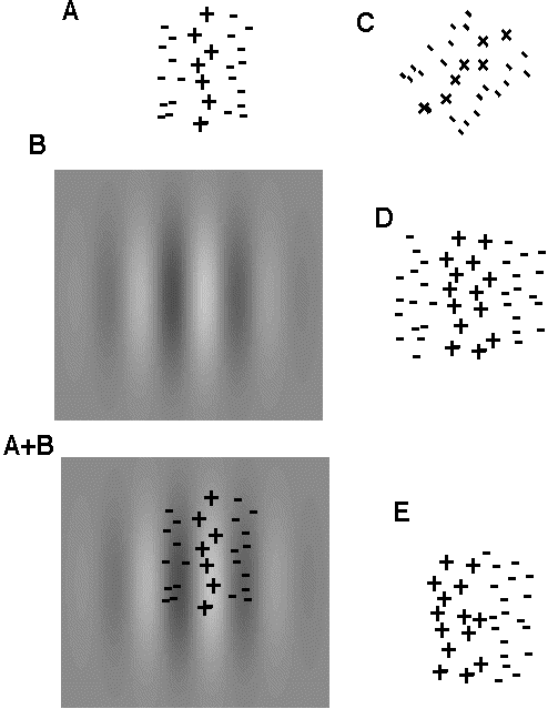
Fig. 1. Receptive fields and sinusoidal grating stimuli.
A. The receptive field of a neuron in the primary visual cortex is the plot of the region of the retina where light spots either excite (+) or inhibit (-) the firing of the cortical neuron.
B. A patch of sinusoidal grating.
A+B. If this sinusoidal patch is shown on the retina so it is centered on the the receptive field shown in A (see the lower left), the corresponding neuron will be strongly excited because the light stripe will stimulate the excitatory subregion (yielding a good deal of excitation) and the black stripes will stimulate the inhibitory subregions (yielding very little inhibition). A good deal of excitation coupled with very little inhibition produces a big response!
C, D, and E. The receptive fields of different cortical neurons differ in orientation (C), the width of their subregions (D), and their symmetry (E is odd symmetric, while A, C, & D are even symmetric).
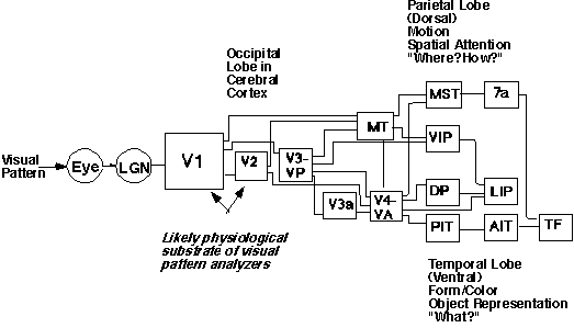
Fig. 2. (from Graham, 1992). A simplified diagram of the visual cortical connections in the macaque monkey with additional labeling added to describe the current idea of at least two major streams of information processing. The lines without arrows imply that connections are reciprocal. The visual system of the human is thought to be rather similar to the macaque. The presumed physiological substrate of the multiple analyzers in the psychophysical model is cortical area V1. Adapted from Movshon, A. (1990) Visual processing of moving images. In Images and Understanding, Barlow, H., Blakemore, C., and Weston-Smith, M. (eds). Cambridge University Press: Cambridge, England.