Purpose. To seek evidence for -- or against -- the idea that retinal processing subserves detection in a probed-sinewave task used to investigate light adaptation.
Methods. Detection threshold was measured for a decremental test probe (13 msec, 1.5 deg with a smooth edge) presented at eight different phases on a 10 deg background, flickering sinusoidally at 1.2 and 9.4 Hz. In the dichoptic condition the probe was presented to one eye and the flickering background was presented to the other eye using a system of mirrors. In the monoptic condition the probe and flickering background were presented to the same eye. A Yes/No procedure was used with interleaved staircases.
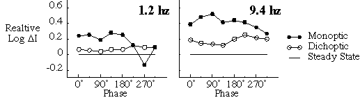
Results. The figure shows relative threshold averaged across four subjects plotted against phase of the flickering background (where 0 deg is the positive zero-crossing). In the monoptic conditions probe threshold (filled symbols) varied as a function of phase. In the dichoptic conditions probe threshold (open symbols) was only slightly elevated relative to the steady state (no symbols), particularly at 1.2 Hz. These results suggest that the processing is primarily retinal. However, the modest modulation of the dichoptic results and the slightly elevation relative to the steady state suggest some cortical contribution, particularly at 9.4 Hz.
Conclusions. These results suggest that this probed-sinewave task taps into primarily retinal processes.