


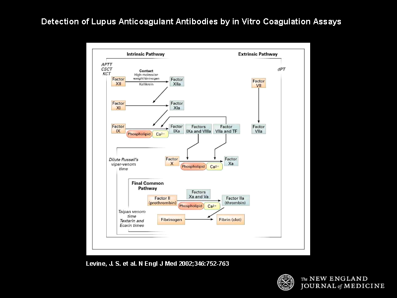
Figure 1. Detection of Lupus Anticoagulant Antibodies by in Vitro Coagulation Assays.
The various coagulation tests used to detect lupus anticoagulant activity are indicated in italics, and the figure shows a simplified schematic diagram of the coagulation pathway evaluated by each of these tests. The coagulation cascade is a result of the enzymatic conversion of each factor (orange boxes) to its activated, or enzymatic, form (blue boxes), which then, in combination with an activated cofactor, catalyzes the subsequent reaction. The intrinsic coagulation pathway is initiated by contact activation on glass, silica, or kaolin (as in the activated partial-thromboplastin time [APTT], colloidal-silica clotting time [CSCT], and kaolin clotting time [KCT] assays), whereas the extrinsic coagulation pathway is initiated by the formation of a complex between tissue factor (TF) and factor VIIa (as in the dilute prothrombin time [dPT] assay). Both the intrinsic and extrinsic pathways result in the conversion of factor X to activated factor X (factor Xa). Finally, both intrinsic and extrinsic pathways converge on the final common pathway, the activation of prothrombin to thrombin followed by the conversion of fibrinogen to fibrin. Russell's viper venom directly activates factor X. Taipan, Textarin, and Ecarin snake-venom extracts directly activate prothrombin but have different cofactor requirements. Taipan venom activation of prothrombin requires phospholipid and calcium but not factor Va. Textarin activation of prothrombin requires phospholipid, calcium, and factor Va, whereas Ecarin activation of prothrombin is independent of cofactors and does not require phospholipid, calcium, or factor Va. The activation of prothrombin to thrombin, like several other reactions in the coagulation cascade, requires the presence of phospholipid and calcium. These phospholipid-dependent reactions are believed to be targeted by lupus anticoagulant antibodies in vitro. Although both the intrinsic and extrinsic pathways are invaluable for understanding clot formation in vitro, the extrinsic pathway has the dominant role in vivo.