


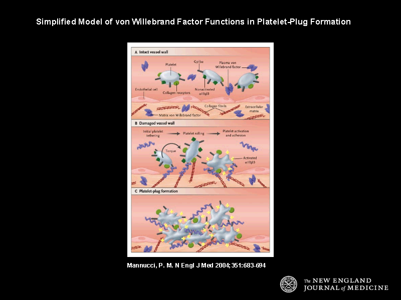
Figure 1. Simplified Model of von Willebrand Factor Functions in Platelet-Plug Formation.
In the intact vessel wall (Panel A), endothelial cells hamper the interactions of circulating platelets and their membrane glycoproteins Ib{alpha} (GpIb{alpha}), nonactivated IIb-IIIa ({alpha}IIb{beta}3), and collagen receptors GpVI and {alpha}2{beta}1 with von Willebrand factor and collagen fibrils localized in the subendothelial extracellular matrix. When the vessel wall is intact and blood flow is normal, plasma von Willebrand factor that is present in a coiled structure and platelets coexist in circulating blood with minimal interactions. In the damaged vessel wall (Panel B), collagen and von Willebrand factor of the subendothelial matrix become exposed to flowing blood and shear forces. Plasma von Willebrand factor efficiently binds to exposed collagen and uncoils its structure, supporting the adhesion of circulating platelets in synergy with collagen. Bound von Willebrand factor interacts, at first, only with the platelet receptor GpIb{alpha} and platelet tethering occurs. This interaction has a fast dissociation rate, and platelets tethered to the vessel wall still move in the direction of flow (rolling). In this interaction, collagen receptors GpVI and {alpha}2{beta}1 bind to collagen and promote platelet adhesion and activation in synergy with the von Willebrand factor-GpIb{alpha} interactions. Once platelets are activated (represented by irregular margins), a conformational change of {alpha}IIb{beta}3 enhances its affinity for the ligand von Willebrand factor (receptors are shown as yellow crosses). This event, together with the rolling of platelets due to the von Willebrand factor-GpIb{alpha} interaction, allows {alpha}IIb{beta}3 to bind platelets to the vessel wall (Panel C); {alpha}IIb{beta}3 is also responsible for platelet-to-platelet interactions that eventually lead to platelet-plug formation mediated by von Willebrand factor and, at slow flow conditions, by fibrinogen (not shown).