

SEEING SMALL:
Visualization in chemistry
EVER SINCE the unveiling of the double helix of DNA, the best-known image of molecular
biology, biochemists and molecular biologists have tried to elucidate and manipulate the
structures of other molecules, such as proteins, with important roles in health and disease. These
molecules are relatively large and complex, but even the largest (about 1 millionth of an inch in
diameter) is outside the range of the most sensitive microscope."Seeing" the atoms inside these molecules involves an earlier visualization technique known as X-ray diffraction, which depends on the principle that an array of atoms will scatter X-rays in a regular pattern onto a photographic plate. These patterns enable the scientist to build the atomic structure of the molecule. For years atomic patterns were drawn by hand, as Max Perutz did in his 22-year effort to unravel the structure of hemoglobin, a protein involved in oxygen transport.
With the rise in computer power, scientists found that computers could visualize the 3-D shape and structure of proteins with great efficiency. What once took X-ray crystallographers months and years could now be done in days. The computers also could rotate the molecule and see how different parts fit together. The practical utility of computer visualization was not lost on pharmaceutical researchers, who for years had been developing new drugs by trial and error. Now they could alter the structure of a biologically active molecule and see how the new entity might behave.
Columbia scientists have written some remarkable software for drug design. The first program, known as MacroModel (to distinguish it from earlier software called Model), came out of the work of the Department of Chemistry's W. Clark Still, a chemical synthesis expert. This model helps in analyzing the structure and behavior of relatively small molecules-not more than 100 atoms. Still says MacroModel was not strictly created as a visualization tool but can be used for that purpose, especially when generating ensembles of molecules.
According to Still, companies use MacroModel in different ways but mainly for "conformation searching." Almost all molecules have multiple conformations, but only a few are biologically active. Determining the common structural threads will help researchers trace the cause of biological activity to the molecular level and enable them to manipulate structures as desired.
Still attributes much of the program's success to its simulation of the molecule in the real world-that is, surrounded by water or another solvent. "Most molecular models," says Still, "have been developed with the molecule as an ideal crystal structure or in a vacuum. I believe ours is the first to model the molecule rapidly in solution, its natural habitat."
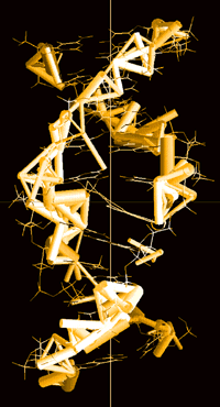 The second type of software developed at
Columbia displays 3-D structural perspectives of large biological molecules such as the proteins,
which range from 2,000 atoms per molecule to 200,000. GRASP (Graphical Representation
and Analysis of Surface Properties) was developed by Anthony Nicholls and Barry Honig at
the Department of Biochemistry and Molecular
Biophysics at P&S. The program enables molecules to undergo visual transformations of
surface and structure. On the screen they range from thin, wiry spaghetti-like configurations to
bulbous conglomerates of atoms, looking like a mass of rock candy. Changes in any of these
structural forms by addition of new molecules can have profound functional consequences.
The second type of software developed at
Columbia displays 3-D structural perspectives of large biological molecules such as the proteins,
which range from 2,000 atoms per molecule to 200,000. GRASP (Graphical Representation
and Analysis of Surface Properties) was developed by Anthony Nicholls and Barry Honig at
the Department of Biochemistry and Molecular
Biophysics at P&S. The program enables molecules to undergo visual transformations of
surface and structure. On the screen they range from thin, wiry spaghetti-like configurations to
bulbous conglomerates of atoms, looking like a mass of rock candy. Changes in any of these
structural forms by addition of new molecules can have profound functional consequences.
The GRASP software can be traced back to Honig's interest in protein electrostatics and his development of computational methods to predict the electrical properties of proteins. Nicholls, who developed GRASP to represent these properties graphically, says it has opened up new ways to think about proteins and how they are modeled. "It isn't enough to make the pictures," he says. "You have to move them around until you get a 3-D shape that you can work with."
Visualization lets researchers pinpoint the operational sites of complex molecules and thus prepare the ground for intervention. Neil McDonald, research fellow in biochemistry, worked for three years elucidating the structure of nerve growth factor, a protein important in trauma recovery and degenerative disease treatment. In subsequent study, Nicholls became intrigued by a large contiguous blue surface at one end of the molecule and speculated that this region was the molecule's binding site to a receptor. Further work revealed that nerve growth factor actually binds to two types of receptors. Research is ongoing and may lead to a means of interfering with binding.
Wayne Hendrickson, a professor of biochemistry and molecular biophysics at P&S, focused on a more powerful visualization technique to decipher structures that otherwise would be impenetrable. Using synchrotron radiation, he solved the structure of human chorionic gonadotropin, as well as the part of the insulin receptor that lies just beneath the cell membrane, the tyrosine kinase domain. The latter discovery has led to the first explanation of how the cell surface signals the nucleus.
Finally, visualization has yielded one of the most important images in immunology: the major histocompatibility complex (MHC) molecule, whose structure has been determined by researchers at Columbia, Harvard, and elsewhere. Their work reveals how a few molecules could account for the body's extraordinary specificity in matching antibody to antigen.
GRASP is currently licensed to 150 companies and is distributed on the Internet.


