SBPMD Histology Laboratory Manual
Epithelium: Micrographs
Examine the electron Micrographs so that you understand the ultrastructural equivalents of the structures you have seen under the microscope.
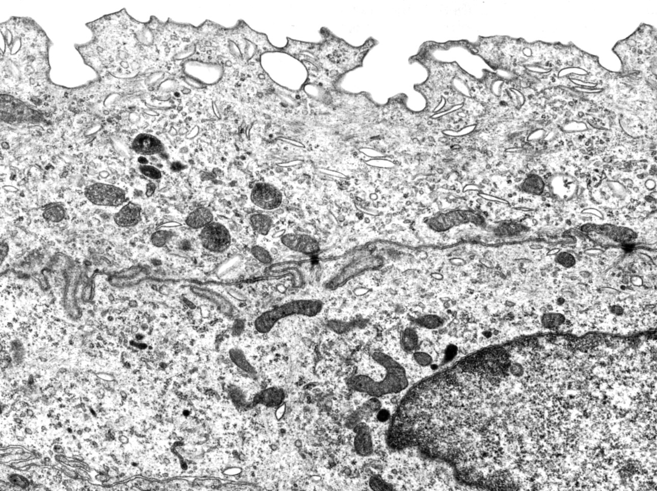
Junctional Complex | |
| Click to see enlarged view | |
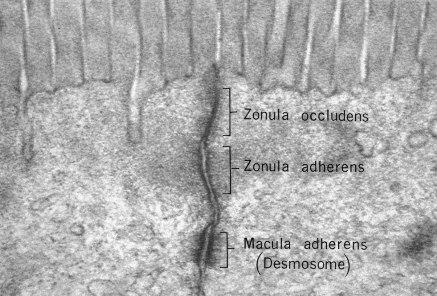 |
Apical region of Intestinal lining (hamster) illustrating the junctional complex. The components are: the zonula occludens (tight junction) formed by two zonula occludens strands from neighboring cells fusing in a zipper like fashion. This restricts the movement of water and other molecules through the intercellular space as well as lipid rafts containing specific proteins within the plasma membrane itself. This establishes polarity across the epithelial lining. The zonula adherens is an anchoring junction that provides lateral adhesion between epithelial cells. The zonula adherens contains transmembrane cell adhesion molecules (CAMs, mainly cadherins) that link the adjacent cells externally. Intracellularly they act as binding sites for actin filaments through vinculin and catenin. The macula adherens (desmosome) provides a localized spotlike junction. It consists of an intracellular plaque with desmoplakins and plakoglobins into which intermediate filaments insert and transmembrane glycoproteins whose extracellular proteins desmocollin and desmoglein (cadherin family) bind identical molecules in the neighboring cell. |
| |
Simple Squamous Epithelium | |
| Click to see enlarged view | |
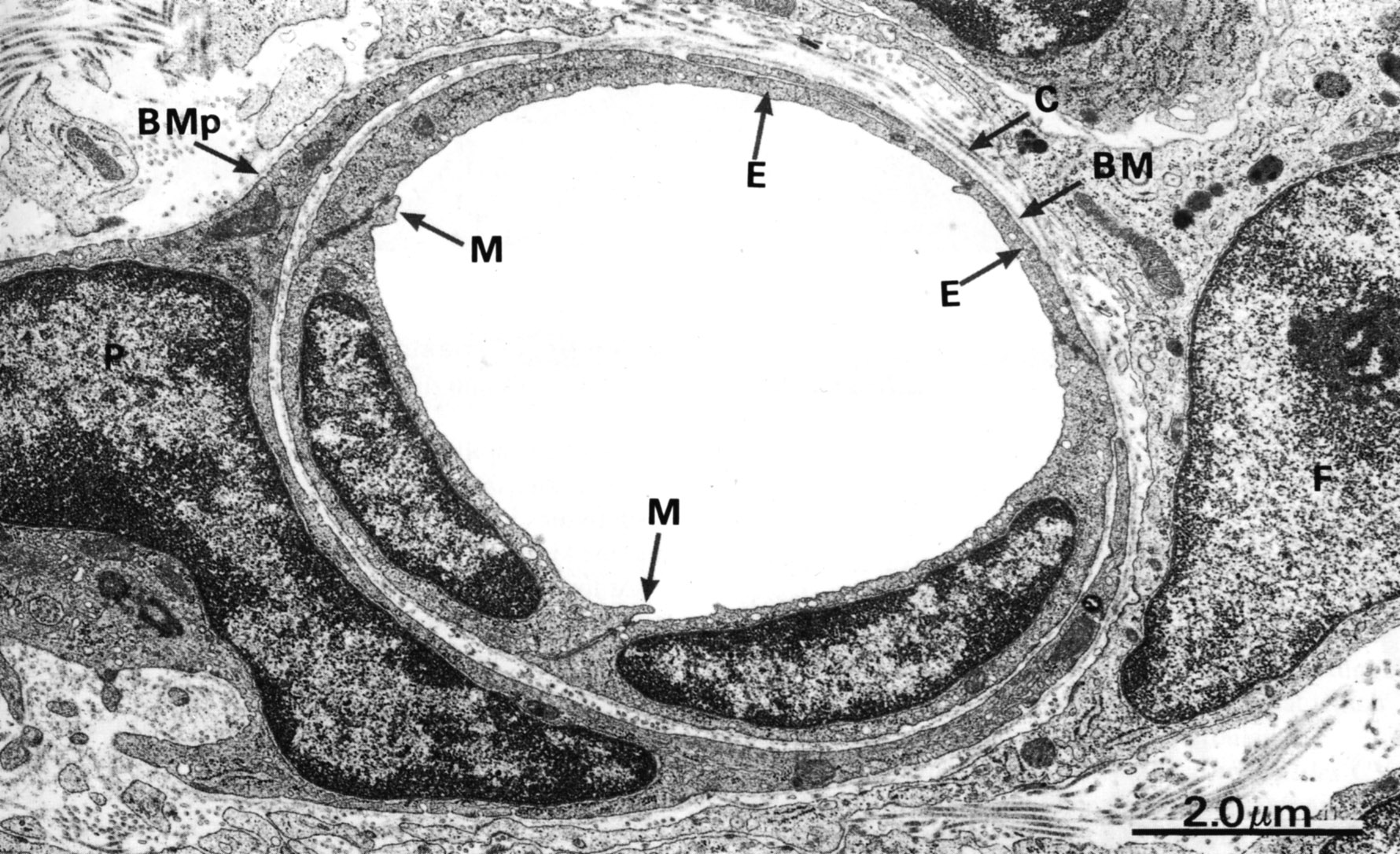 |
The lining of blood vessels is simple squamous epithelium, termed endothelium. A capillary is illustrated. In this unusual section portions of 4 endothelial cells have been cut. The highly attenuated cells are connected by tight junctions. Outside of the basement membrane (BM) there is collagen (C ). Pericytes (P) are associated with the capillary. (M) indicates small cytoplasmic marginal folds. |
| |
Simple Cuboidal Epithelium | |
| Click to see enlarged view | |
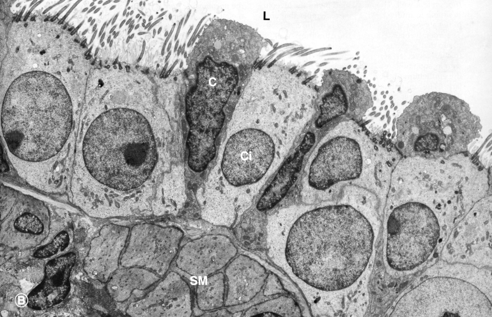 |
Terminal bronchiole showing ciliated cells (ci) and secretory cells (Clara cells, C). Lumen (L), smooth muscle, cut in cross section (SM) |
| |
Simple Columnar Epithelium | |
| Click to see enlarged view | |
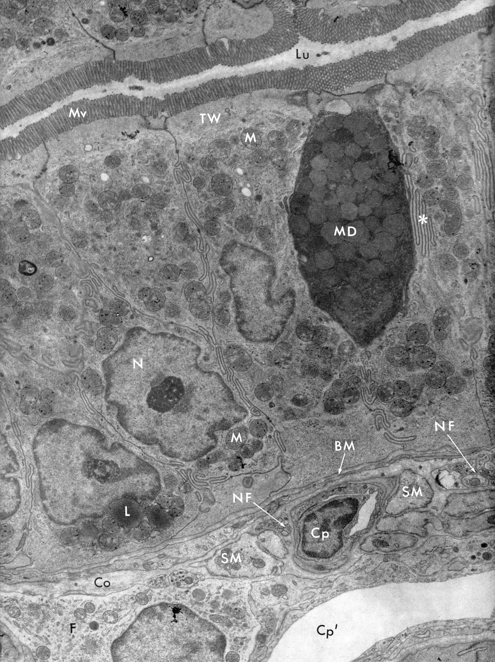 |
This simple columnar intestinal epithelium is made up of enterocytes (the majority) and goblet cells. The enterocytes have a microvillous border (Mv) facing the lumen (Lu). The terminal web (TW) is a meshwork of actin filament, intermediate filaments and spectrin in the apical cytoplasm. The lateral membranes of adjacent cells are highly tortuous*. The goblet cell contains mucus droplets (MD), Nucleus (N), basement membrane(SM), nerve fibers (NF), fibroblasts (F), collagen fibers (Co) lipid droplet (L), mitochondria (M), capillary (Cp). |
| |
Pseudostratified epithelium, Trachea (human) | |
| Click to see enlarged view | |
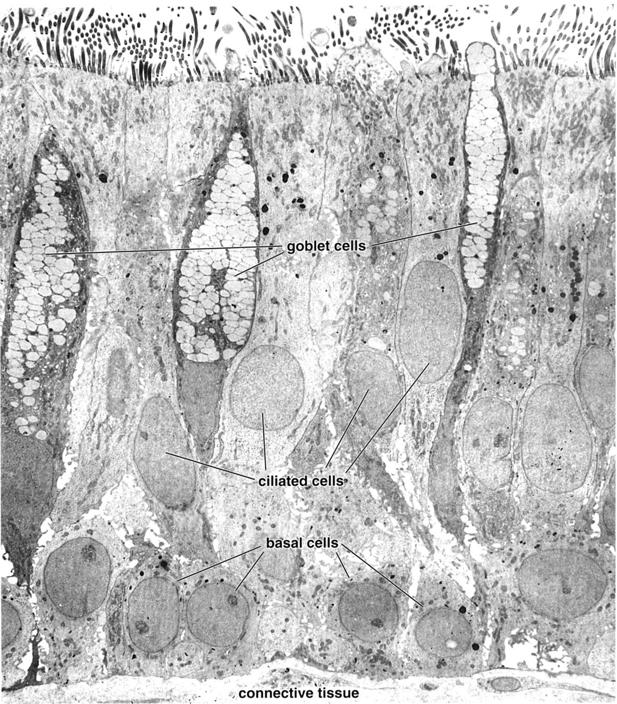 |
There are three main cell types in this pseudostratified epithelium, all of which lie on the basement membrane: ciliated cells that reach the lumen; goblet cells with mucinogen granule which also reach the lumen; and basal cells which are confined to the basal portion of the epithelium (and act as progenitors for the other types). |
| |
Transitional Epithelium (urothelium) | |
| Click to see enlarged view | |
 |
The superficial layer of transitional epithelium is shown including the cytoplasm of one luminal lining cell. Its plasma membrane contains plaques that are hinged such that when the bladder is contracted, peripheral membrane is internalized, forming vesicles. These are unfolded when the bladder is distended. |