SBPMD Histology Laboratory Manual
Central Nervous System: Brain
The central nervous system consists of the brain and spinal cord.
#86 Cerebellum, Human
Open with WebViewer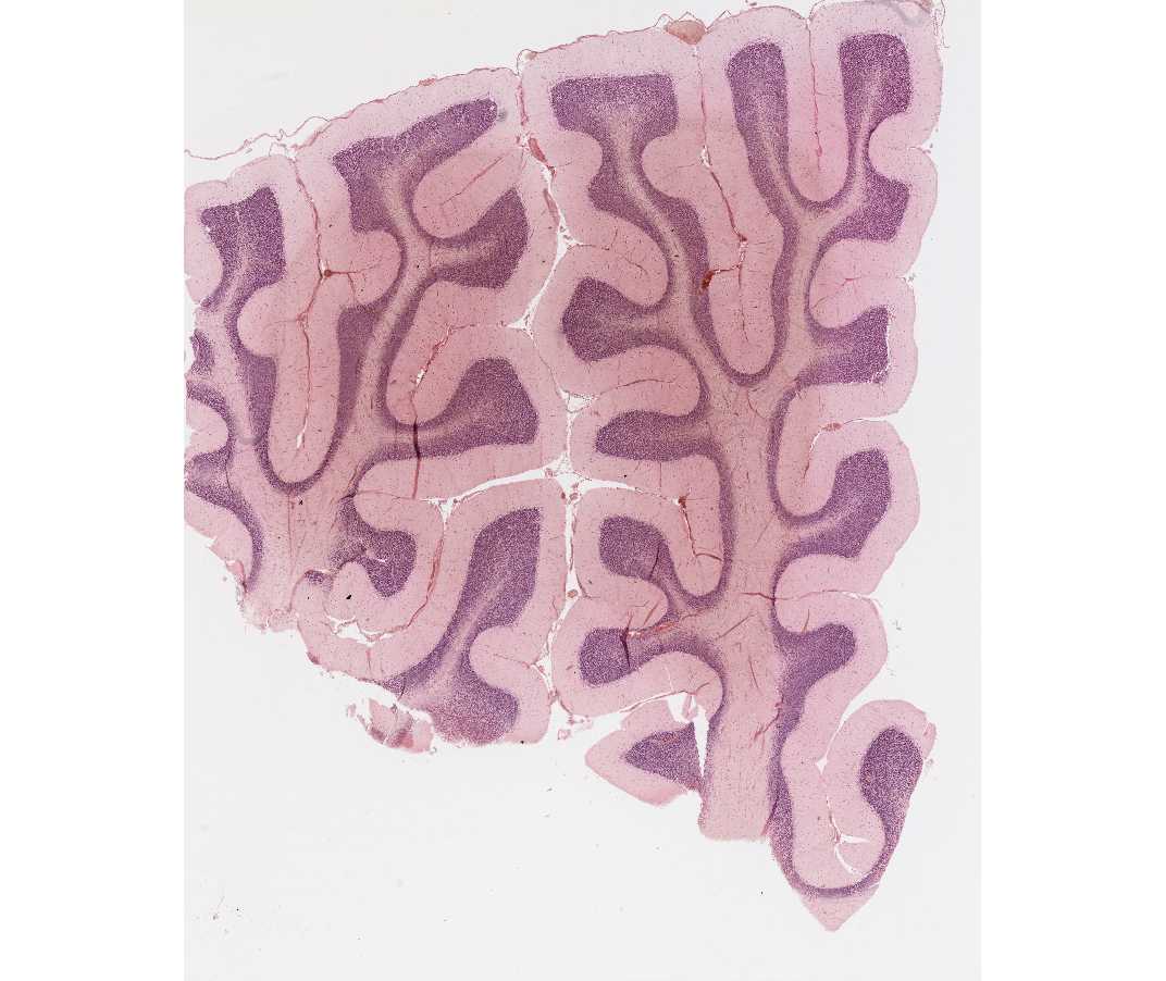
With the naked eye, observe the pale staining branches of the central white matter (medulla), surrounded by a darkly stained cortex. With the scanning objective, identify the outer, pale-staining molecular layer of the cerebellar cortex, and the inner, basophilic granular layer of the cortex. Both the molecular layer and granular layer constitute the gray matter. The molecular layer contains axons and dendrites, but relatively few neurons compared to the granular layer.
A word of caution: On these sections of the cerebellum, the cut surfaces may result in the exposure of the pale-staining medulla (white matter) at the surface of the section, where it could be confused with the molecular layer of the cortex. Try to find a surface covered by the meninges, to insure that you are indeed looking at the cortical surface.
With the 10x objective, examine the junctional zone between the molecular and granular layers of the cortex. Note the large, flask- shaped cells aligned in a row; these are the cell bodies of Purkinje cells.
The basophilic nuclei of the granular layer, which superficially resemble lymphocyte nuclei, belong to granule cells. Axons of these cells (not visible with H&E) extend into the molecular layer and relay neural information to this layer.
Demonstration Slide: Cerebellum (developing mouse)

An immunocytochemical demonstration of the Purkinje cells of the cerebellum can be seen on this slide. The section was treated with a rabbit antibody to calcium binding protein (Calbindin) (primary antibody). It was then incubated with goat anti-rabbit immunoglobulin (secondary antibody). Sites of antigen/antibody complexes were visualized with DAB that forms a brown precipitate that can be seen in the cell body and the neuronal fiber processes.
#108 Cerebral Cortex, Cat, Nissl
Open with WebViewer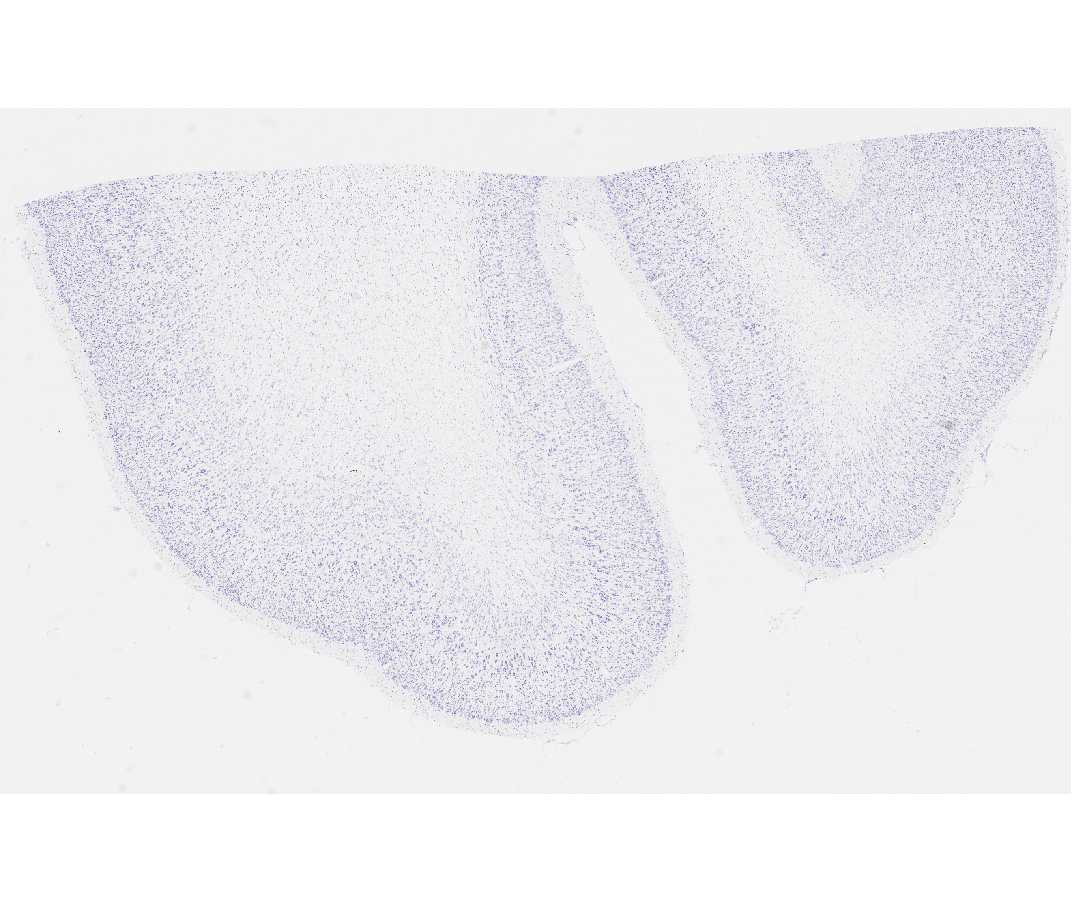
With the scanning objective, find the gray matter (cerebral cortex) and white matter, which in this stain can be identified by the rows of small oligodendrocyte and astrocyte nuclei between the unstained axons and their myelin sheaths. The cortex, itself, is divided into 6 layers, not all of which are clearly distinguishable in this slide. (Do not try to find all of them.) At higher power study the large pyramidal cells, which are prominent in deeper parts of the cortex. Study the nucleus, nucleolus, and Nissl substance at higher power. Note the similarity of the large pyramidal cells to the large motor neurons in the ventral horn of the spinal cord.
#111 Cerebral Cortex, Cat, Golgi, 100µm, Celloidin embedded
Open with WebViewer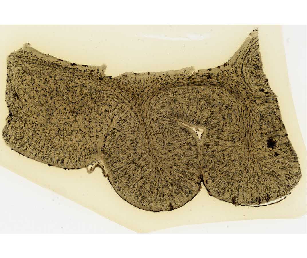
The Golgi procedure results in 1-2% of neurons impregnated with heavy metals. No one is sure why some but not all cells are stained. This preparation shows the detailed architecture of individual neurons. The thickness of the sections allows one to appreciate the amount of volume occupied by a neuron's dendritic tree.