SBPMD Histology Laboratory Manual
Connective Tissue: Dense, Regularly Arranged
Collagenous
#3 Muscle and Tendon, Human, H&E
Open with WebViewer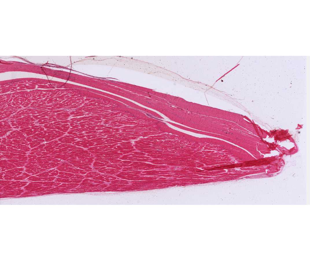
The thick, collagenous bundles of the tendon run parallel to each other, slight waviness of the tissue is due to fixation. Rows of fibroblasts with heterochromatic nuclei are aligned between the collagenous bundles. Compare the appearance of the collagen bundles (Type I collagen) and fibroblasts with that of the skeletal muscle fibers on the same section.
Note: Inspect the example in the demonstration microscope if your slide is not good
#11 Bone, rib (H&E)
Open with WebViewer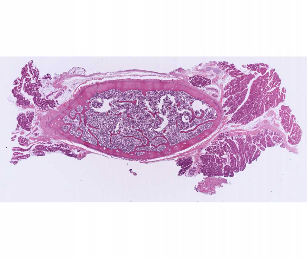
Find the regions of the dense fibrous regularly arranged connective tissue (tendon). Collagen is stained pink and can be distinguished from skeletal muscle that is stained purple. Note the fibroblasts aligned along the collagen fibers in the tendon. These are flattened cells with heterochromatic nuclei.
Elastic
#20 Aorta, Rabbit, (Weigert stain)
Open with WebViewer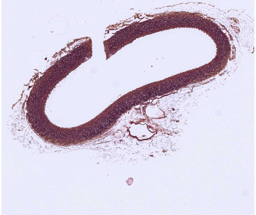
The fibers are predominantly elastic rather than collagenous. Elastic fibers stain reddish-brown to black and form prominent fenestrated, elastic sheets in the aorta. The elastic tissue in the aorta s made by smooth muscle cells.
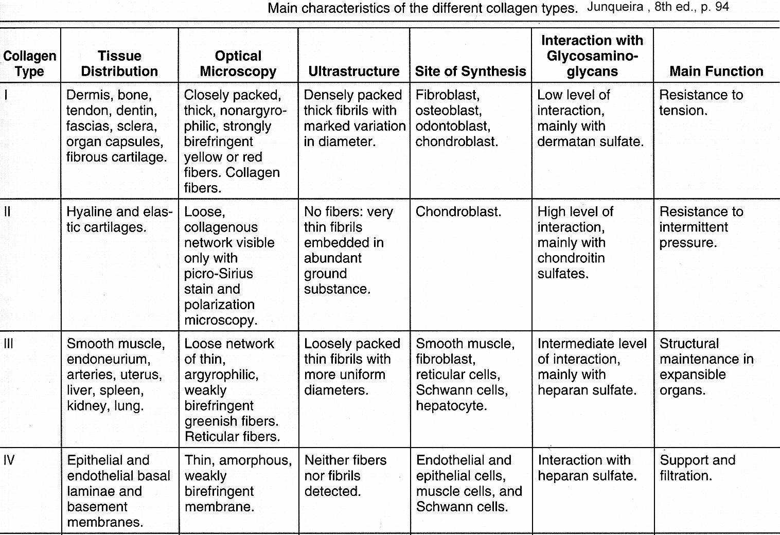
The collagen types listed below are the most common.