SBPMD Histology Laboratory Manual
Cartilage
Cartilage is a specialized type of connective tissue whose unique combination of rigidity, elasticity and resilience is due primarily to the special properties of its matrix. As in other connective tissues, its matrix is composed of fibers (collagenous or elastic) and a ground substance that is rich in extracellular glycosaminoglycans (particularly the chondroitin sulfates). Cartilage is avascular, but its matrix is permeable to tissue fluids carrying nutrients and waste products.
Cartilage is the primary skeletal tissue of the fetus, and it serves as a model for the development of endochrondral bone. In the adult, cartilage is more restricted in its distribution, but it forms the articular surfaces of diarthrodial joints, the skeleton of the external ear and the septum of the nose, supporting rings and plates of the trachea and bronchi, and intervertebral discs. Three types of cartilage are found in the adult; these are classified according to the predominant component of their extracellular matrix. As in other connective tissue classifications, there are gradations between these basic types.
Mesenchyme & Cartilage Development
#91, 92 Pig Embryos, 15mm and 25mm
Locate the neural tube and below it, the notochord. In slide #91 the mesenchymal cells surrounding the notochord have condensed and are beginning to differentiate into cartilage cells. These cells will eventually form the vertebrae. In later stages (#92), the cartilage cells become separated by the deposition of collagen and secretion of matrix into the extracellular space. (Note: Few slide boxes will have three embryo slides but most should have two of the stages. If you are missing slides, look at your neighbors'.)
Hyaline Cartilage
#5 Trachea
Open with WebViewer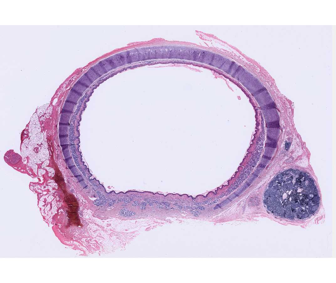
With the naked eye and with the scanning lens, locate the incomplete rings of hyaline cartilage in the wall of the trachea. At higher magnification observe that a perichondrium surrounds the cartilage; this merges with the cartilage on one side and with the surrounding connective tissue of the other side. Blood vessels within the perichondrium provide the blood supply for the avascular cartilage. Chondroblasts are cells adjacent to the perichondrium and recently derived from it. They are not yet completely embedded in the matrix. Mature cartilage cells or chondrocytes are surrounded by matrix and lie within spaces called lacunae. In life the chondrocytes completely fill the lacunae.
Note that the cartilage matrix is relatively homogeneous and basophilic. This is due to the masking of the collagen fibers by the high concentration of the glycosaminoglycans in the ground substance. The matrix immediately surrounding the lacunae is more intensely basophilic. This zone is the territorial matrix.
Why is the territorial matrix more intensely basophilic, and what is its significance? Note that the region outside the territorial matrix may be less basophilic in places. What is the most likely reason for this?
Elastic Cartilage
#6 External Ear (Weigert Elastic Stain)
Open with WebViewer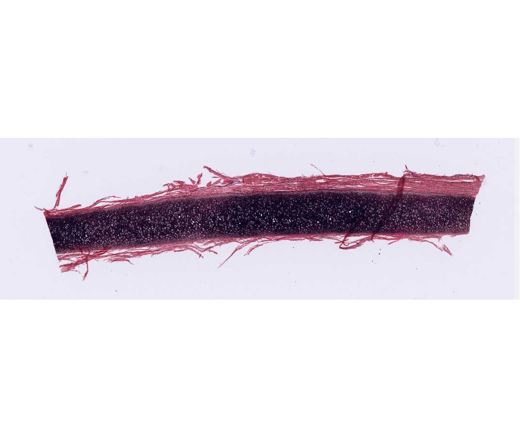
Elastic cartilage provides support with flexibility. The general organization of this type of cartilage is similar to that of hyaline cartilage, except that elastic fibers predominate over collagen fibers in the matrix. Elastic fibers are stained specifically (black) by the Weigert's stain. Where else does elastic cartilage occur in the body?
Questions
- What are the mechanisms of cartilage growth?
- What is the distribution of blood vessels in cartilage and how does this relate to the nutrition of cartilage?