SBPMD Histology Laboratory Manual
Cardiovascular System: The Heart
Illustrations of the heart in your gross anatomy textbook or atlas will help you to orient your slide and to locate the atrium, ventricle and mitral valve.
#17 Heart, Monkey, Sagittal Section (Mallory-Azan)
Open with WebViewer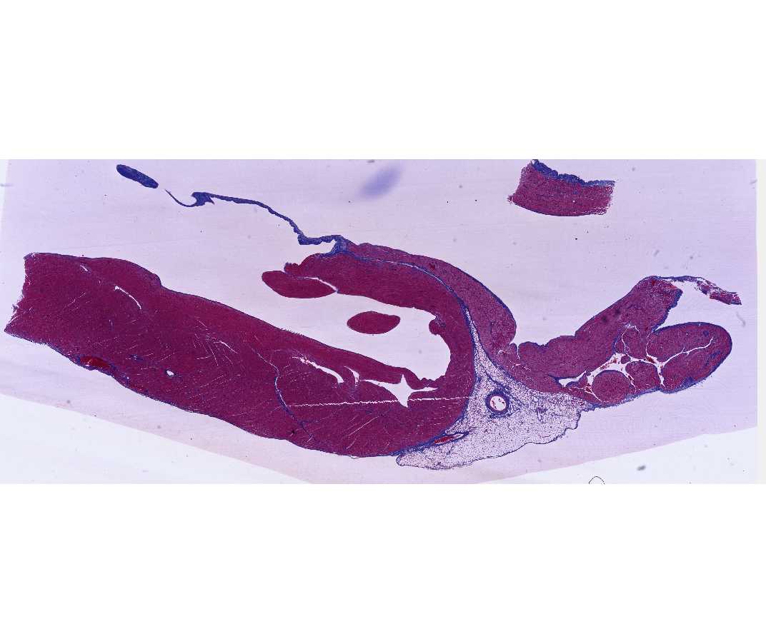
The epicardium in the region of the atrium contains fatty connective tissue and vessels of the coronary circulation. This can be seen by gross inspection of the slide, and it serves as a landmark for microscopic study. Note the blue-staining collagen fibers of the fibrous trigone between the atrium and ventricle. The distribution of blue-staining collagen fibers reveals the fascicle organization of the myocardium. In areas where muscle fascicles are longitudinally sectioned, one can see intercalated discs that appear as red-staining bars or step-like lines perpendicular to the long axis of the fiber.
The heart valves are extensions of the endocardium. They contain a connective tissue core and an endothelium on their free surface. The structure of the arteries and veins of the coronary circulation should also be studied.
#18 Heart and Mitral Valve, Human Sagittal Section (Masson's Trichrome) (Not scanned)
This slide is similar in organization to that of #17; however, certain cytological details are not as clear, and it is difficult to identify intercalated discs in this preparation. Note the connective tissue that comprises the mitral valve.
#19 Heart, Ventricle, Calf (PTAH Stain)
Open with WebViewer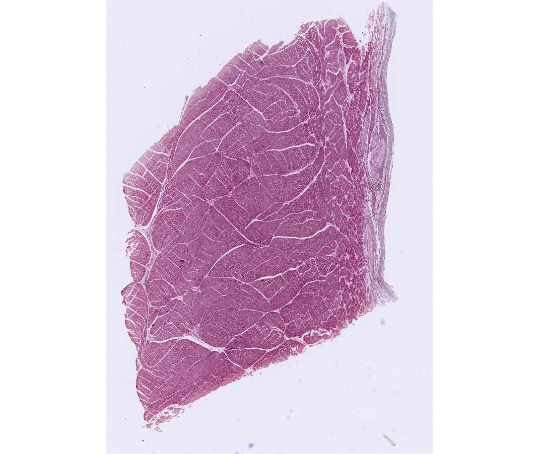
Purkinje fibers are branches of the impulse conducting system of the heart; in particular, they are branches of the A-V bundle of His. Purkinje fibers are hypertrophied cardiac muscle fibers that are specialized for conducting an impulse rather than for contraction. They contain one or two nuclei, centrally situated in a pale staining mass of sarcoplasm that is rich in mitochondria and glycogen. The Purkinje fibers of the cow are especially large, and are excellent for the study of the conducting system. Individual muscle fibers are grouped in fascicles that are sectioned in both cross and longitudinal section. The fascicles are bounded by collagenous connective tissue containing blood vessels of the coronary circulation, sympathetic and parasympathetic nerve fibers, and terminal branches of the A-V bundle of His. Major branches of the A-V bundle lie outside the myocardium in the subendocardium. Purkinje fibers in the myocardium are encapsulated by connective tissue except in areas where the terminal branches merge into muscle fascicles. This is seen in favorable longitudinal sections as a point where the Purkinje fibers become smaller, more densely stained and become indistinguishable from fascicles of muscle fibers.