SBPMD Histology Laboratory Manual
Cardiovascular System: Micrographs
Examine the electron Micrographs so that you understand the ultrastructural equivalents of the structures you have seen under the microscope.
Cardiac Muscle | |
| Click to see enlarged view | |
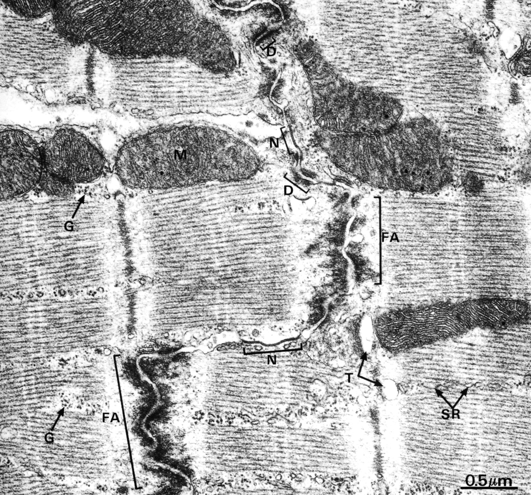 |
Cardiac muscle cells are joined by the interdigitating junction known as the intercalated disk. It consists of three types of membrane-to-membrane contact. The predominant type of contact is the fascia adherens (FA) in which actin filaments at the ends of terminal sarcomeres insert into the fasciae adherentes and thereby transmit contractile forces from cell to cell. Desmosomes (D) provide anchorage for intermediate filaments. Gap (nexus) junctions (N) are present mainly in the longitudinal portions of the interdigitations. These are important for coordinating function among the cardiac muscle cells. There are numerous mitochondria (M) and abundant glycogen (G). Profiles of the sarcoplasmic reticulum (SR) and parts of T tubules (T) can be identified. |
| |
Types of Capillaries | |
| Click to see enlarged view | |
 |
Three basic types of blood capillaries are illustrated. They are differentiated by the continuity of the endothelial cell and the basal lamina. A, continuous capillary; b, fenestrated capillary; c, discontinuous capillary (sinusoid). Rat diaphragm, pancreas and liver, respectively. |
| |
Continuous Capillary (myocardium, cat) | |
| Click to see enlarged view | |
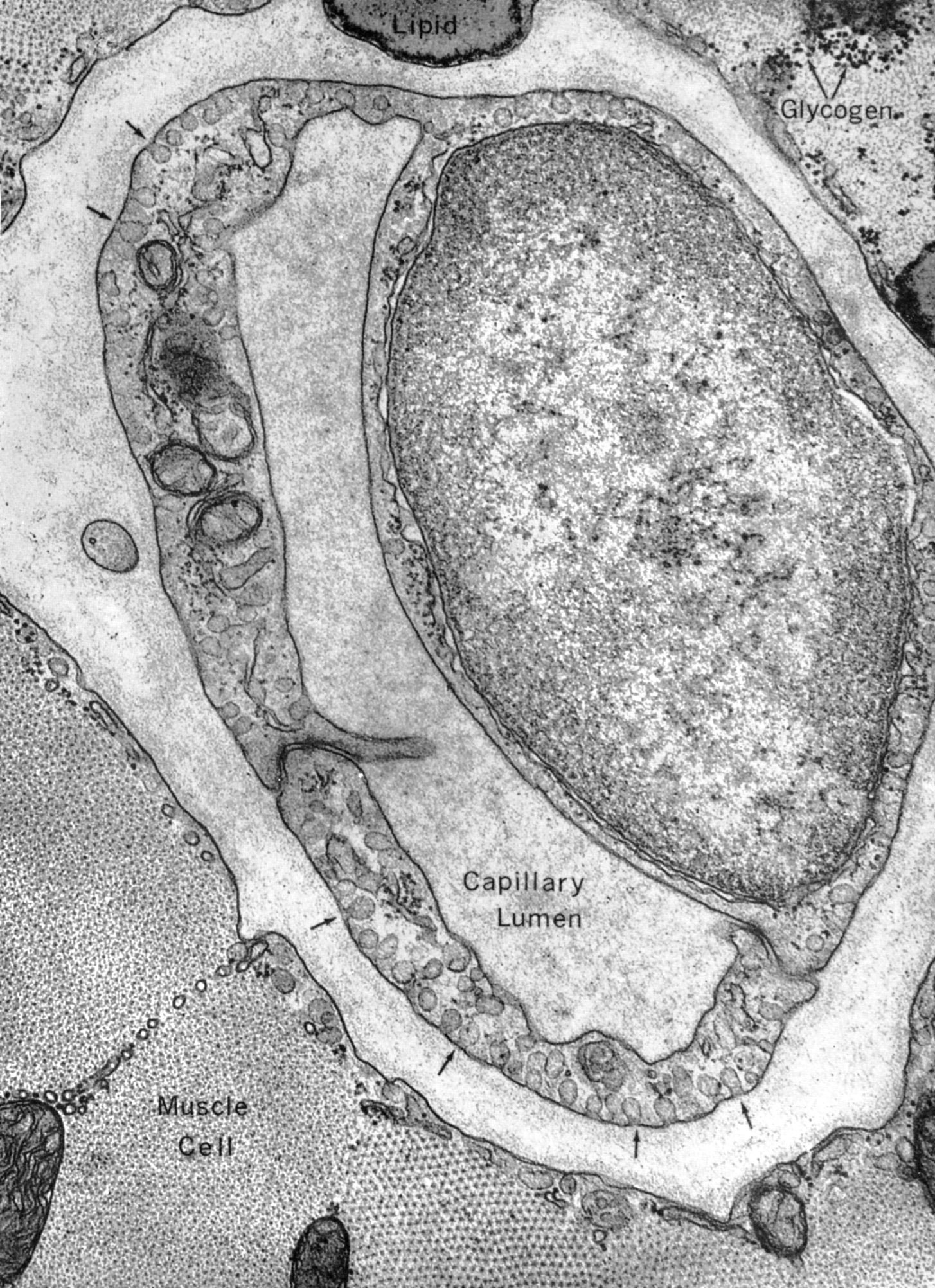 |
Endothelial cells of continuous capillaries are joined by tight junctions. They contain pinocytotic vesicles (arrows). There is a continuous basal lamina.
|
| |
Fenestrated Capillary (nonglomerular region of kidney) | |
| Click to see enlarged view | |
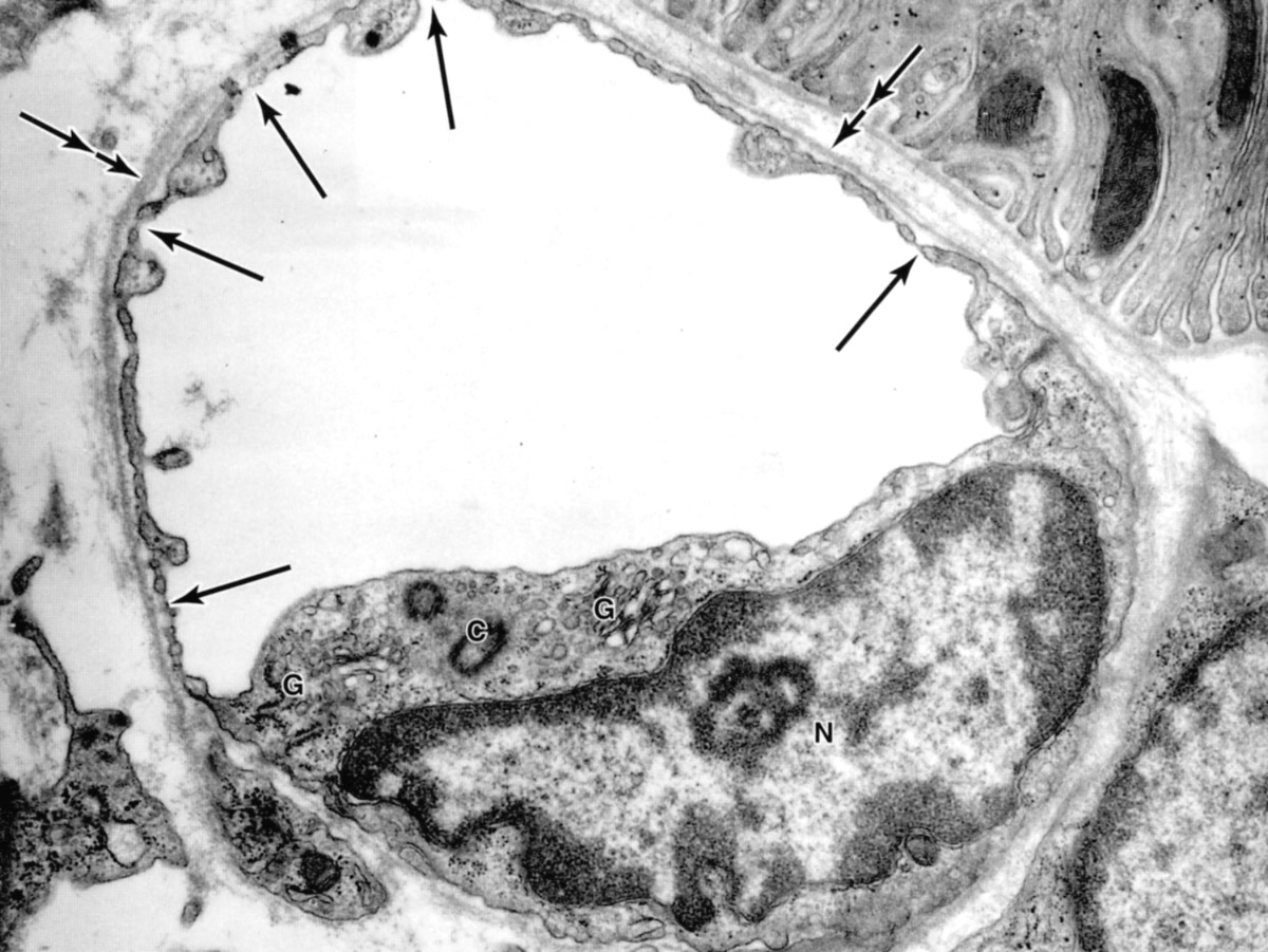 |
Arrows indicate fenestrae closed by diaphragms. In this cell the nucleus (N), Golgi complex (G), and centrioles (C ) can be seen. Note the continuous basal lamina on the outer surface of the endothelial cell (double arrows). |
| |
Sinusoidal Capillary | |
| Click to see enlarged view | |
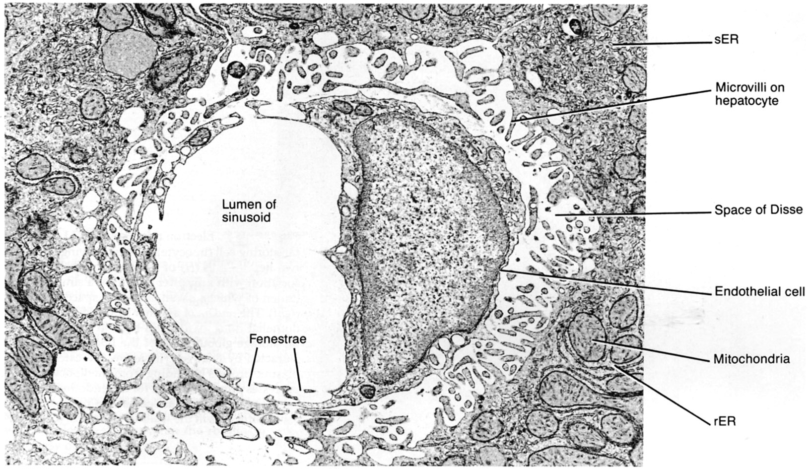 |
Liver sinusoid in cross section (rat). Open fenestrae are evident in the endothelial cell cytoplasm. The space of Disse is between the sinusoidal wall and the hepatocytes. |
| |