SBPMD Histology Laboratory Manual
Blood, Hematopoiesis: Micrographs
Examine the electron Micrographs so that you understand the ultrastructural equivalents of the structures you have seen under the microscope.
Eosinophil (rat bone marrow) | |
| Click to see enlarged view | |
 |
Note bilobed nucleus (N). The specific granules (Gr) are the most conspicuous constituent of the cytoplasm in light microscopy, but electron microscopy reveals the presence of lysosomes as well. The specific granules are discs (seen here in both cross * and longitudinal section) that contain a crystalloid body which is responsible for the refractivity of the granules in light microscopy. They contain an arginine-rich protein called major basic protein. There are elements of rough endoplasmic reticulum (ER) and ribosomes. |
| |
Megakaryocyte | |
| Click to see enlarged view | |
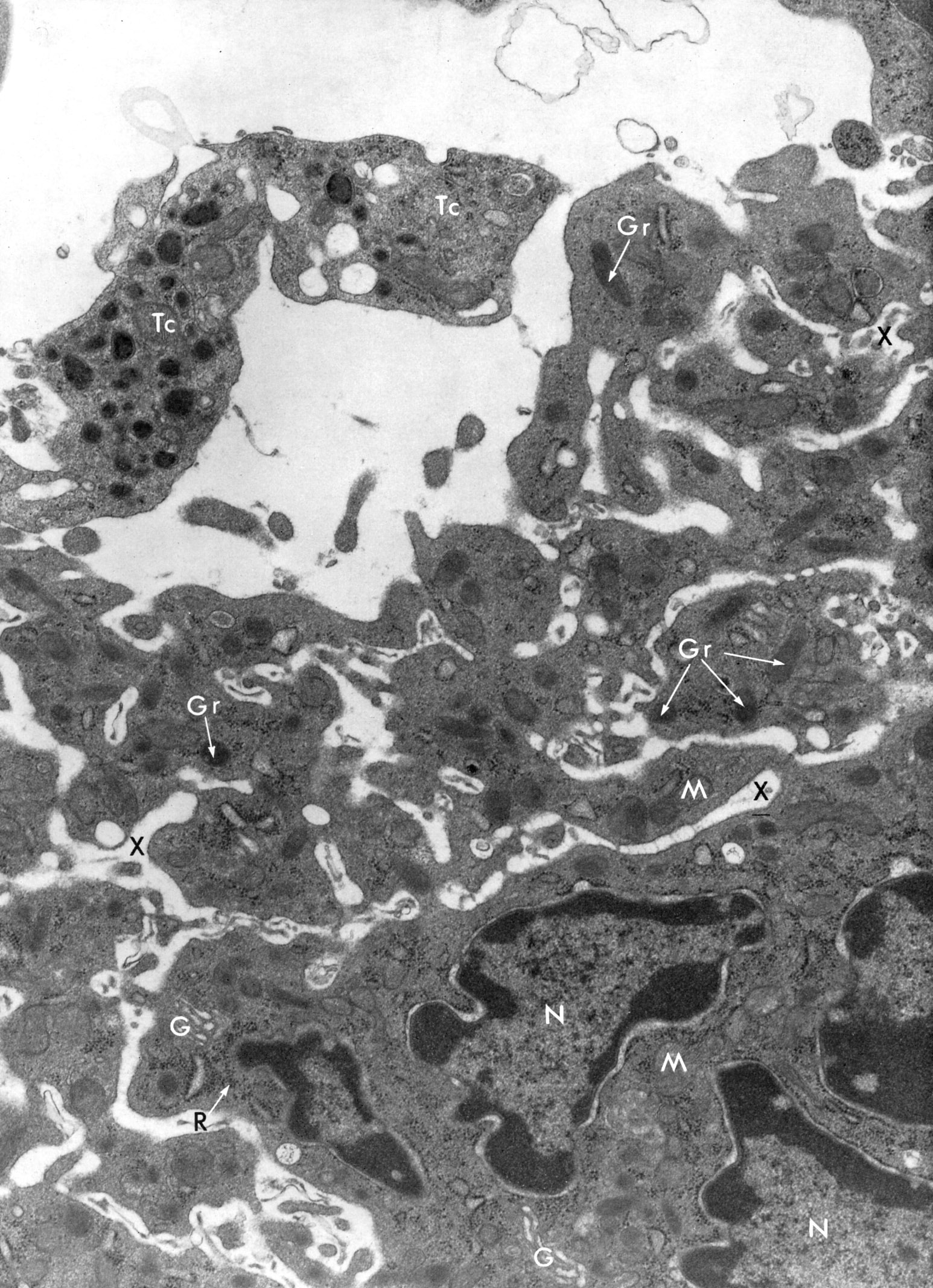 |
Portion of a megakaryocyte (bone marrow rat) showing a part of the single nucleus (N) which is multilobular and polyploid. The cytoplasm contains mitochondria (M), elements of Golgi (G), ribosomes (R), rough endoplasmic reticulum and granules (Gr). Individual platelets (upper left) are devoid of nuclei but contain mitochondria, ribosomes, rough endoplasmic reticulum and granules (platelet at left is labeled, Tc). Demarcation zones (X) are sites where platelets are derived by fragmentation. |
| |