SBPMD Histology Laboratory Manual
Lymphatic Tissues: Lymph Nodes
#22 Lymph node, Dog, Reticular Tissue (Silver Stain, Bielschowsky
(If you do not have this slide in your box, please borrow it from your neighbor.)
Open with WebViewer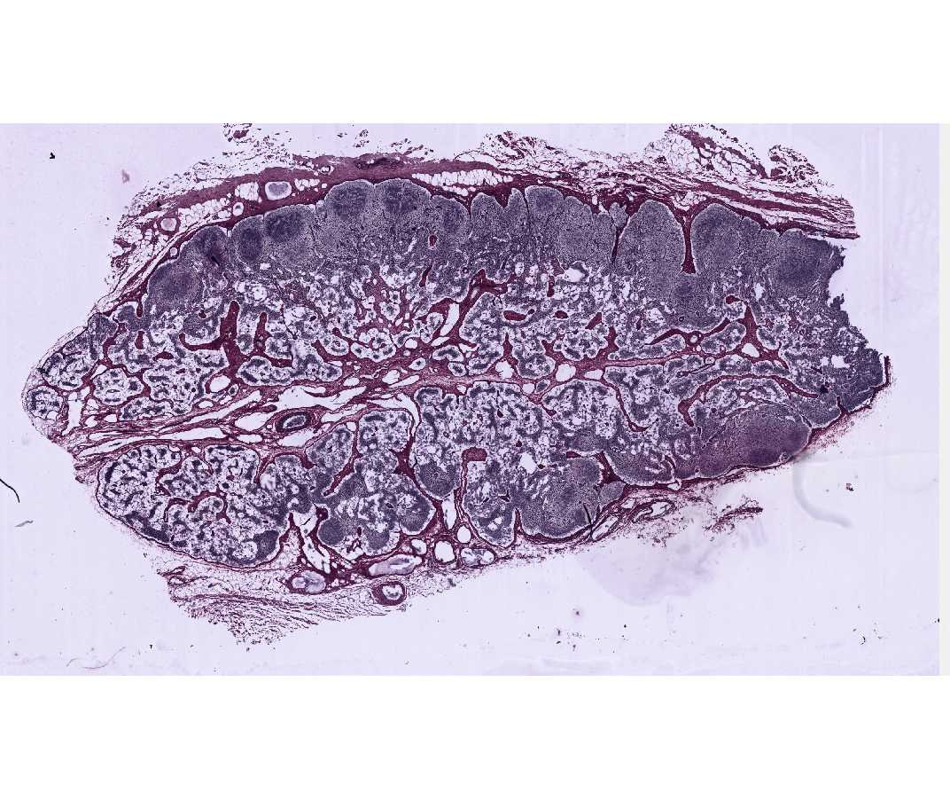 In lymph nodes (and spleen and other regions that contain cells that produce antibodies), the lymphoid tissue consists of two principal elements: (1) a "fixed" or stable element (stroma) represented by a fine three dimensional meshwork of large branched reticular cells, which is reinforced by a similar meshwork of argyrophilic reticular fibers, and (2) "free" or unattached and mobile cells, such as lymphocytes and plasma cells, which accumulate within the interstices of the reticular meshwork.
The histological topography of a lymph node is well shown in this preparation although the silver is staining only the reticular stroma, but not the cellular elements. The reticular fibers are selectively stained black by the silver reticulum stain and are arranged in loose spidery meshworks. Type I collagen is stained reddish brown.
Identify the following structures:
1. The connective tissue capsule stained red by the counterstain and covered on its external surface by loose connective tissue in which there are fat cells, blood vessels and some afferent lymphatic vessels with delicate valves.
2. the subcapsular (or marginal) sinus, lying immediately below the capsule.
3. small cortical sinuses (trabecular sinuses) on either side of the trabeculae.
4. connective tissue trabeculae, stained red and extending from the capsule into the cortical region.
5. the cortex, composed of lymphatic tissue with nodules (that are poorly defined in this node since there has been no recent antigenic or infectious stimulation).
6. The thymus-dependent area comprises the mid- and deep-cortex, i.e., the non-nodular region of the cortex (paracortex).
7. the medulla, characterized by lymphatic tissue arranged in branching and anastomosing medullary cords and medullary sinuses.
8. an indented region, the hilus (or hilum), which is surrounded by the medulla and contains connective tissue, a few fat cells, blood vessels, nerve bundles and large efferent lymphatic vessels with delicate valves (to prevent the backup of lymph into the node. Your section may not pass directly through the center of the hilus so you may not see the efferent lymphatic vessel exiting the gland.
Be sure you understand the cellular interactions and activities within both the cortical and medullary regions of the lymph node.
#31 Bronchial lymph node, Monkey (H & Azure II-Eosin)
Open with WebViewer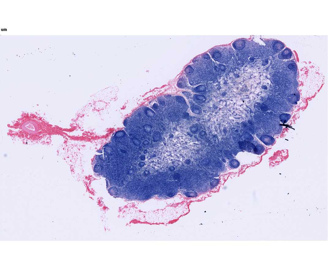
This is a good slide for studying the vascularization and cytological features of the different regions of the node. Under low power identify the cortex, the non-nodular thymus- dependent area and the medulla. In the medulla and identify the region of the hilus, if present. Notice the arteries, veins, and efferent lymphatic vessels. Under low power in the cortex examine the germinal centers and the dark surrounding zone within the lymphatic nodules. The dark zone surrounding the germinal center is composed of densely packed small and medium-sized lymphocytes, separated from each other by layers of flattened pale-staining reticular cells. Under high magnification and then oil, the light zone (germinal center) of the follicle may be observed to contain abundant lymphocytes. Note the plasmablasts which are large cells with a large nucleus and prominent nucleolus; the nucleus is more or less centrally located in the cell and is surrounded by a relatively narrow rim of strongly basophilic cytoplasm. Some plasmablasts in mitosis are evident in this zone. There are also a few free macrophages with engulfed cellular debris. Some pale-staining nuclei of reticular cells can also be seen.
Within the thymus-dependent area of the cortex, identify the postcapillary venules lined by unusual cuboidal endothelial cells. Notice the numerous small lymphocytes traversing the walls of these venules. The postcapillary venules are the site of entrance of B and T lymphocytes into the parenchyma of the lymph node. The T cells remain in the thymic dependent cortical zone and the B cells migrate to the nodular regions. Postcapillary venules are not present in the nodular region of the cortex or in the medullary cords.
Next, examine the medullary region of the lymph node. Lymphocytes are seen in the medullary sinuses, which are discontinuously lined by endothelial cells. The medullary cords are more cellular and consist of reticular cells, lymphocytes and a number of plasmablasts and plasma cells. Plasma cells are smaller than plasmablasts. They have abundant, very basophilic cytoplasm, a prominent Golgi zone and an eccentric nucleus whose chromatin has a "cartwheel" appearance. The plasma cell secretes antibody.
#23 Lymph node, Human or monkey (H&E) (not scanned)
Compare this preparation with the preceding slide. The subcapsular sinus is visible below the capsule. The dark lymphatic nodules may be seen to show lighter-staining germinal centers. The presence of prominent germinal centers indicates that the lymph node has received a recent antigenic stimulation. The non-nodular region of the cortex has a less dense concentration of lymphocytes than the nodular region and represents the thymus-dependent area of the lymph node.
#24 Lymph node. Cat injected with India ink through lymphatic system
Open with WebViewer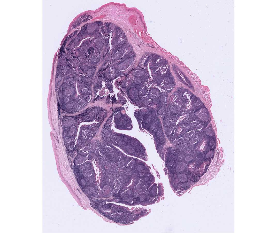
Under low power, notice that the India ink particles are concentrated mainly in the sinuses of the node and to a lesser extent in the medullary cords, where most of the phagocytic cells are located. The ink is virtually absent in the cortical nodules. At higher magnifications, the ink particles may be seen to be contained within the fixed or free macrophages. Clinically the subcapsular sinus (which is only partly injected) is important because it is the first place where groups of cancer cells (metastatic growths) are trapped and multiply in the lymph node. At the hilus of the node, identify adipose tissue, blood vessels and nerve bundles. On the convex surface of the node, opposite the hilus, afferent lymphatic vessels with valves and India ink particles may be seen.