SBPMD Histology Laboratory Manual
Lymphoid Tissue
Examine the electron Micrographs so that you understand the ultrastructural equivalents of the structures you have seen under the microscope.
Plasma Cell | |
| Click to see enlarged view | |
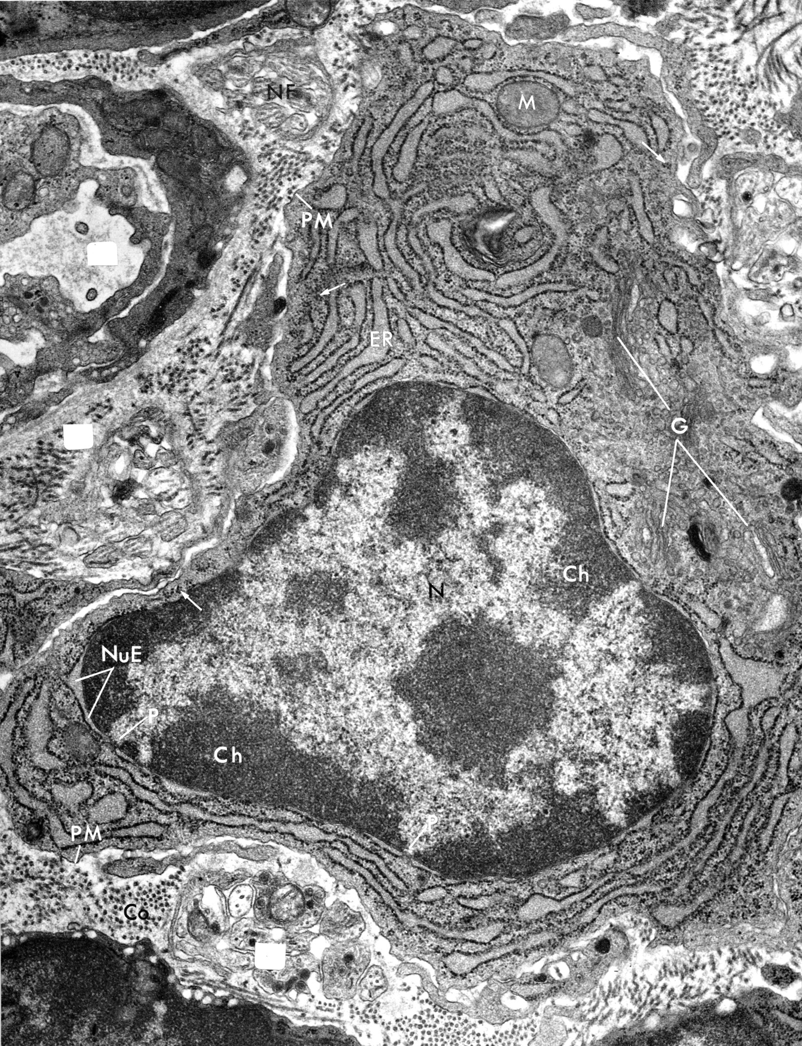 |
Plasma cell from submucosa of a rat. Note the clumps of peripheral heterochromatin (ch) alternating with clear areas of euchromatin and large central nucleolus in the nucleus (N). The zones of euchromatin extend to the pores (P) in the nuclear envelope (NuE), while the heterochromatin is confined to the interpore areas. There is extensive RER and the Golgi region (G ) is well developed. This cells is largely transcribing and processing a single immunoglobulin. (PM, plasma membrane). |
| |
Red Pulp of Spleen | |
| Click to see enlarged view | |
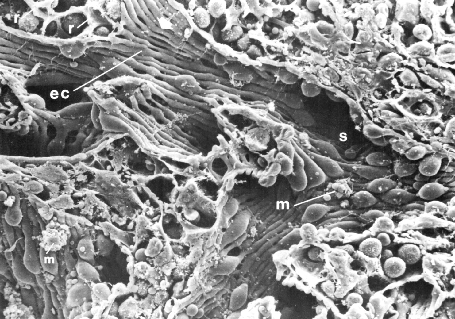 |
Scanning electron micrograph of red pulp of spleen. Sinusoids (s) are lined by elongated endothelial cells (ec). There are macrophages (m) both within the cords and in the sinusoids. There are also reticular cells (r) within the cords. |
| |