SBPMD Histology Laboratory Manual
Lymphatic Tissues: Tonsils
#27 Palatine tonsil, Human. H & E
Open with WebViewer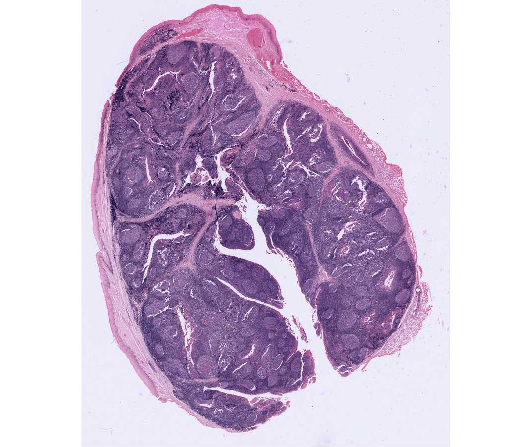
The lymphatic tissue of the tonsillar ring, which is located near the entrance of the throat and which consists of the palatine tonsil (commonly known as "the tonsil"), the pharyngeal tonsil (commonly known as "adenoids"), and the lingual tonsil (on the posterior surface of the tongue). These are unencapsulated lymphatic tissue.
This is a section through a surgically removed palatine tonsil. With the scanning objective, notice the stratified squamous non-keratinized epithelium covering the free oropharyngeal surface of the tonsil. In the underlying lamina propria, identify simple and branched epithelial crypts, sectioned in different planes and representing tubular invaginations of the surface epithelium. The lining epithelium of the crypts may show evidence of keratinization or erosion and can be obscured when heavily infiltrated with lymphocytes. The lumen of some crypts may be seen to contain large numbers of lymphocytes, desquamated epithelial cells and cellular debris. Between the crypts identify the masses of lymphoid tissue containing numerous individual lymphoid nodules. Some of the nodules may merge. Some nodules contain a large pale-staining germinal center. These are secondary nodules. Identify the connective tissue septa that extend at intervals between the crypts and divide the tonsil into lobules, each with an individual crypt as an axis. At one side of the section in the submucosa, note the presence of a pure mucous gland.