SBPMD Histology Laboratory Manual
Respiratory System: Micrographs
Examine the electron Micrographs so that you understand the ultrastructural equivalents of the structures you have seen under the microscope.
Trachea (human) | |
| Click to see enlarged view | |
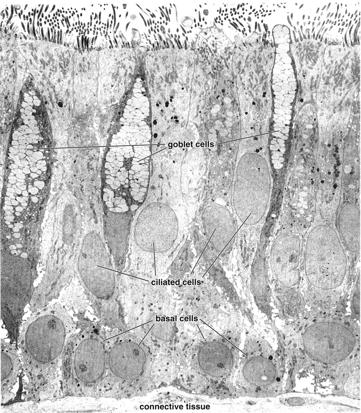 |
The lining of the respiratory tract from the trachea to the larger diameter bronchioles is pseudostratfied epithelium. There are three main cell types in this epithelium: ciliated cells that reach the lumen; goblet cells with mucinogen granules which also reach the lumen; and basal cells which are confined to the basal portion of the epithelium (and act as progenitors for the other types). |
| |
Tracheal lining | |
| Click to see enlarged view | |
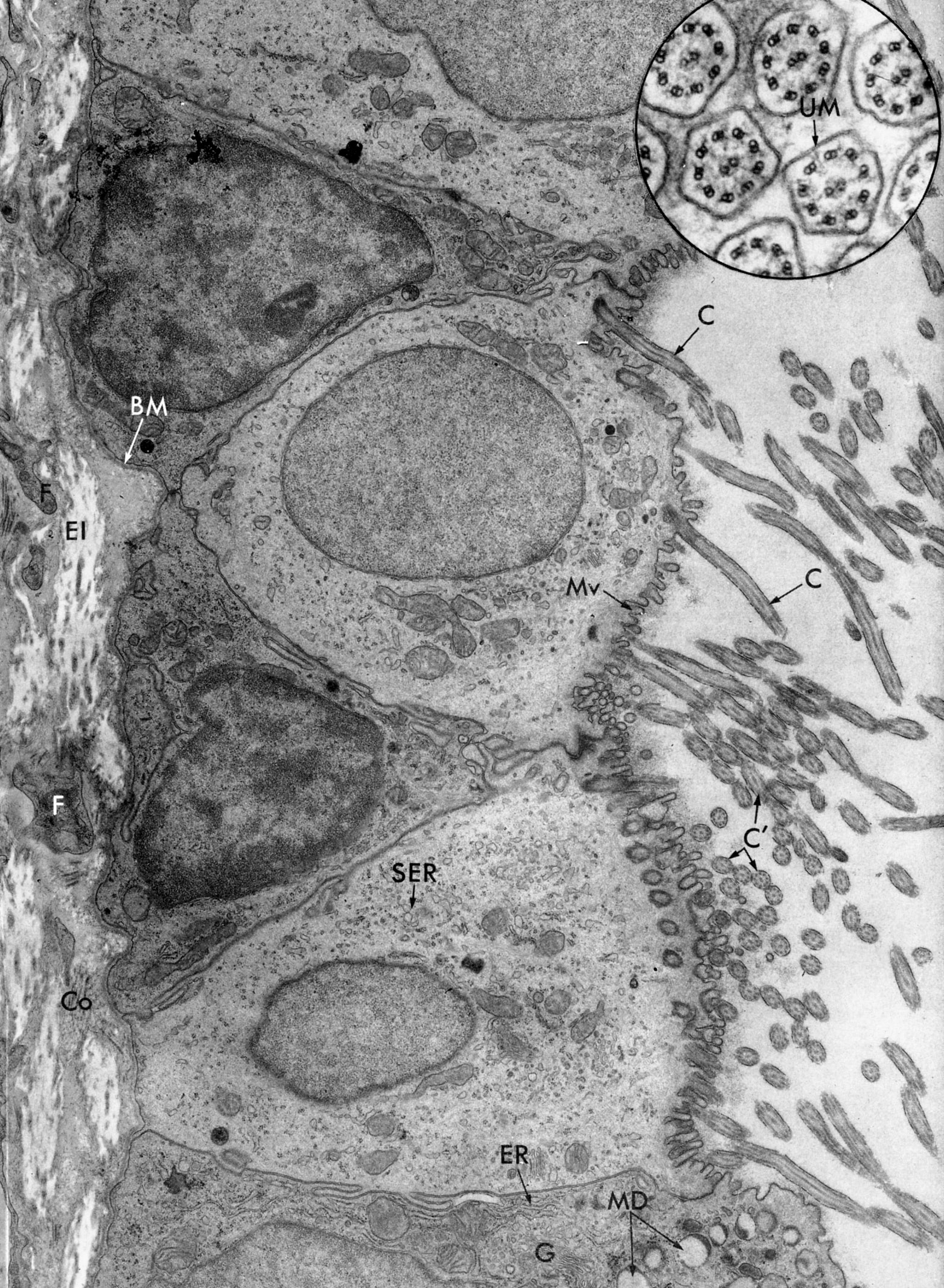 |
Pseudostratified epithelium of trachea of bat (much simpler than human) to illustrate that basal cells, goblet cells and ciliated cells all lie on the basal lamina (BL); but the basal cells do not reach the lumen. The ciliated cells have both microvilli (MV) and cilia, shown here cut in both longitudinal (C ) and cross sections (C). Inset is cross sections of cilia (unit membrane ,UM). The goblet cell (bottom of micrograph) contains secretion droplets (MD) and organelles associated with elaboration of protein for export: (ER) and Golgi (G). The cytoplasm of the non-secretory ciliated cells contains scant Golgi and mainly smooth endoplasmic reticulum (SER). Fibroblasts (F), collagen (Co), elastic fibers (El). |
| |
Ciliated Epithelium with Microvilli | |
| Click to see enlarged view | |
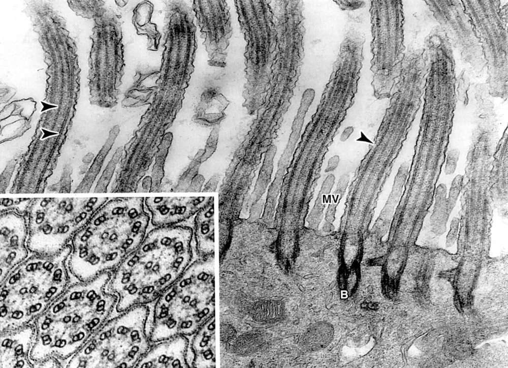 |
Apical portion of ciliated epithelium. Arrows (from left to right) indicate: central, peripheral microtubules of axoneme and plasma membrane. Microvilli (Mv) are also present. Inset is cilia in cross section: Each axoneme contains nine peripheral pairs and two central pairs of microtubules. |
| |
Terminal Bronchiole | |
| Click to see enlarged view | |
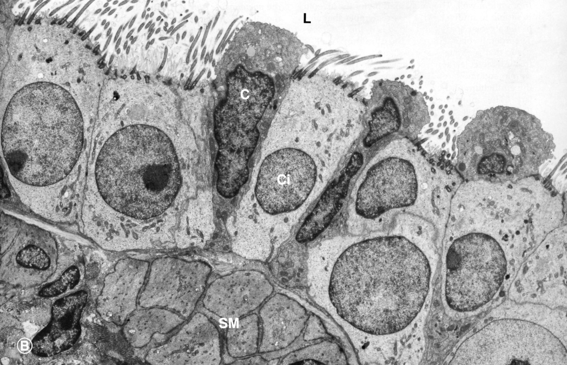 |
Terminal bronchiole showing ciliated cells (ci) and Clara cells (C). Lumen (L), smooth muscle, cut in cross section (SM) |
| |
Alveolus | |
| Click to see enlarged view | |
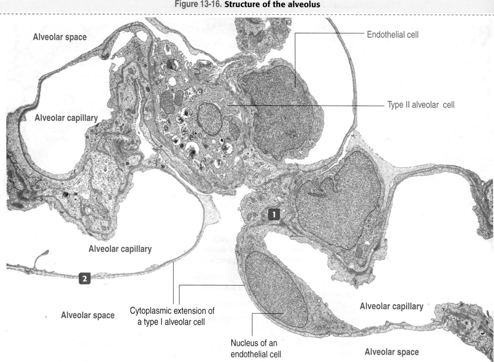 |
The interstitium at (1) is tissue between two layers of alveolar epithelial cells containing elastic and collagen fibers produced by fibroblasts (also known as septal cells). There is no connective tissue over the capillaries (2). Kierszenbaum, AL Histology and Cell Biology 2nd ed., Mosby Elsevier, 2007, p. 388. |
| |
Alveolar Septum | |
| Click to see enlarged view | |
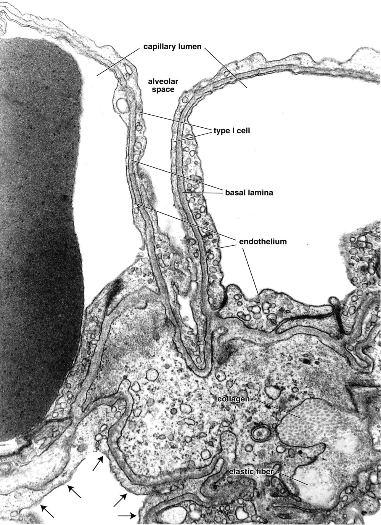 |
The upper part of the micrograph illustrates the thin portion of air-blood barrier where it consists of type I alveolar cells, capillary endothelium and the fused basal lamina shared by both cells. In the thick portion the type I alveolar cell (arrows in lower part of micrograph) rests on basal lamina, and on the opposite side there is connective tissue in which collagen fibrils and elastic fibers are evident. Note erythrocyte (large dark body) in capillary. Ross MH and Pawlina W, Histology, 5th ed., Lippincott Williams & Wilkins, Baltimore, 2006, p. 631. |
| |
Type II Pneumocyte | |
| Click to see enlarged view | |
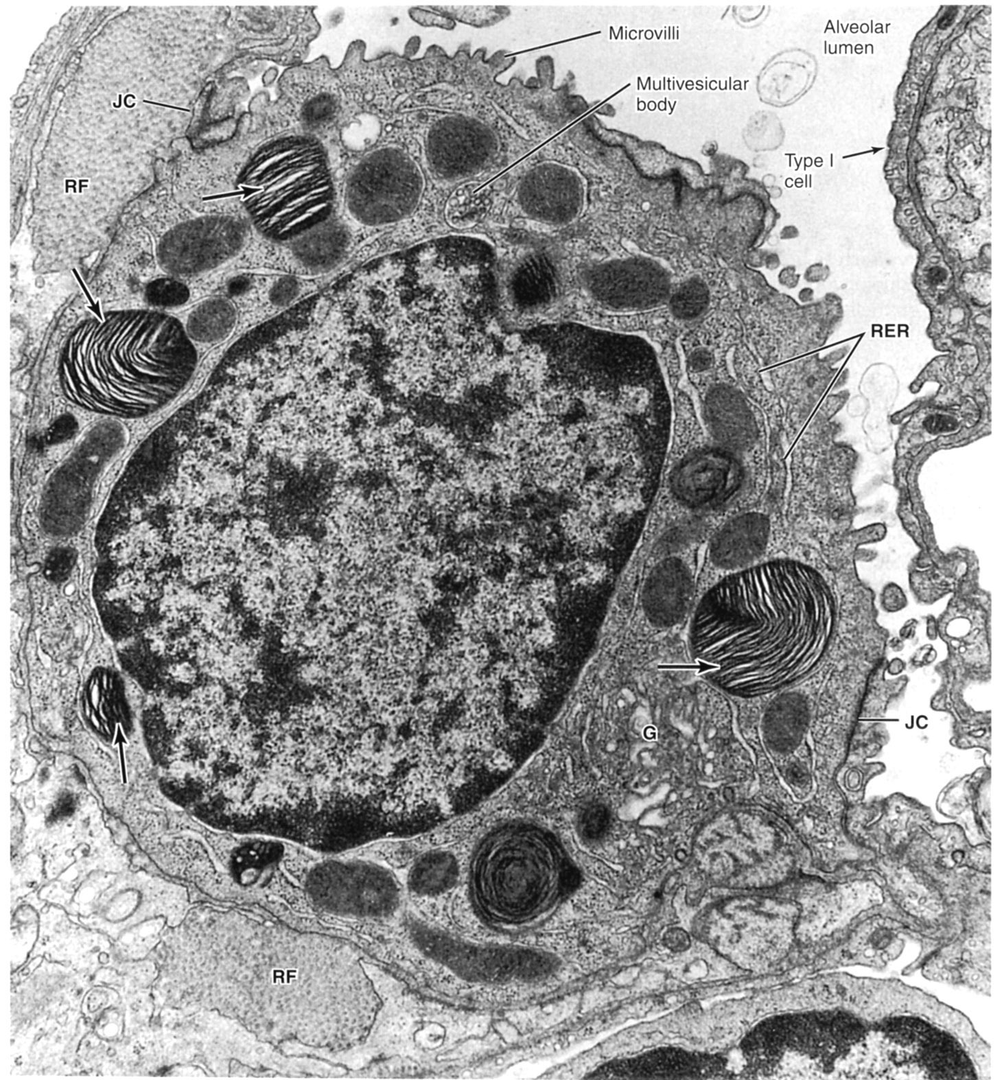 |
Type II pneumocyte protruding into alveolar lumen. Arrows indicate lamellar bodies containing the phospholipids that when released spread over the alveolar surface where they combine with other carbohydrate- and protein-containing secretory products (some of which are derived from Clara cells) to overcome the effects of surface tension which would otherwise cause the alveolar walls to adhere. This allows for normal inflation of the alveoli at birth and for the reinflation of alveoli which collapse after airway obstruction. Rough endoplasmic reticulum (RER), Golgi (G), reticular fibers (RF). Note the microvilli of the type II cell and junctional complexes (JC) with type I cell. |