SBPMD Histology Laboratory Manual
Skin
#4 Skin, thick skin, volar surface, monkey H&E
Open with WebViewer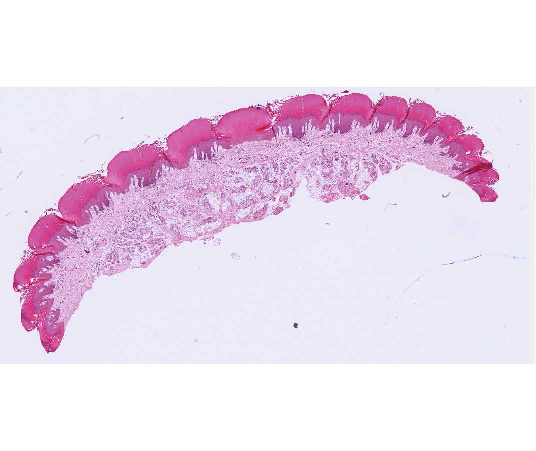
Epidermis: The stratified squamous keratinizing epithelium of the epidermis is made up primarily of keratinocytes. The form and function of these cells changes as they pass from basal to superficial locations. This layer is particularly conspicuous in thick skin. Identify:
Stratum basale (germinativum). Cells of all the layers are generated from the keratinocytes in this layer. Therefore, you may see mitotic figures. The keratinocytes in this and the overlying layers contain melanin granules that have been transferred to them by melanocytes. Cells within the stratum basale with unstained cytoplasm are likely to be melanocytes. Special staining methods are required to identify melanocytes definitively.
Stratum spinosum. This is several cell layers in thickness. The cells are attached to each other by intercellular bridges (desmosomes). Because the cells are artifactually pulled apart, the attachment sites give the cells a spiny appearance.
The cells of the stratum granulosum are recognized by their basophilic keratohyalin granules. This epidermal layer marks the boundary between the non-keratinized and keratinized layers of the epidermis. The stratum lucidum is not clearly identifiable in this slide.
The superficial keratinized layer is the stratum corneum. In some areas it is partially disrupted by sectioning; this is a common artifact that may fortuitously show the lamellar nature of this layer.
Dermis: Papillary layer: loose connective tissue underlying the basal layer of the epidermis, and containing blood vessels, nerves and lymphatic vessels. Dermal papillae may contain sensory nerve endings, the Meissner’s corpuscles. Reticular layer: dense, irregularly arranged connective tissue.
Eccrine sweat glands are present in high concentration in the dermis and subcutaneous tissue. They are coiled tubular glands surrounded by connective tissue. The coiled portion of each gland contains a secretory segment and part of the duct system.
The secretory tubules have a larger diameter and an acidophilic margin, which corresponds to the layer of myoepithelium. The duct tubules are smaller, darker, and formed by a double layer of cuboidal epithelial cells, which may not be obvious in every sectioned duct. A straight portion of the duct passes through the superficial dermis to the basal aspect of the epidermis where it again assumes a coiled pathway. Note the terminal corkscrew-like path of the duct in the stratum corneum.
Pacinian corpuscles are another type of nerve endings, which may be found in some slides in the dermis or subcutaneous tissue.
Subcutaneous tissue (hypodermis): This is loose connective tissue containing abundant adipose tissue. It is a good region to examine glands, ducts, blood vessels and nerves.
#46 Skin, scalp, human H&E.
Open with WebViewer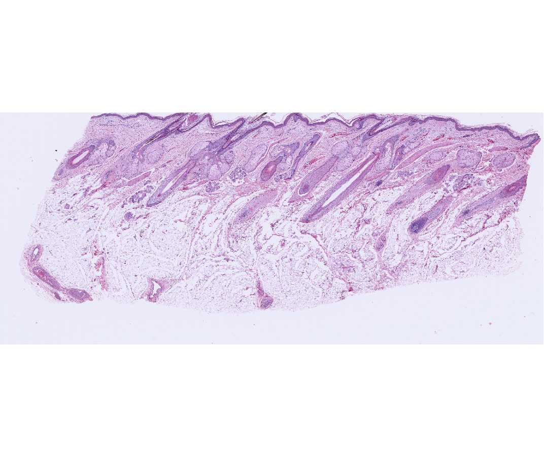
Hair follicles, sectioned longitudinally, are well demonstrated in this slide. The thin epidermis, characteristic of hairy regions, has a lacy or frayed stratum corneum whose appearance is an artifact of sectioning. In life, this layer of the epidermis would be more compact and only the most superficial keratinized cells would be desquamating.
The deepest part of the hair follicle is expanded into a bulb and is invaginated by connective tissue, the dermal papilla. The follicle cells adjacent to the papilla are the germinative cells, and it is by their division and differentiation that a hair shaft and a multi-layered inner root sheath are formed. Melanocytes are adjacent to the dermal papilla and contribute melanin granules to the developing hair. The cytoplasm of melanocytes is unstained. The fully formed hair is usually lost from the section.
Sebaceous glands are associated with the hair follicle and their secretions empty into the hair follicle, between the hair shaft and the follicle wall. Arrector pili muscle is a band of smooth muscle that inserts on the hair follicle, deep to the sebaceous glands. Superficially it inserts at the base of the epidermis. Some eccrine sweat glands are dispersed between the hair follicles.