SBPMD Histology Laboratory Manual
Gastrointestinal System II: Liver
The liver is the largest gland in the body, and it has both an endocrine and exocrine function. The liver is organized into lobes, which are subdivided into lobules. Each liver lobe is surrounded by a thick connective tissue capsule, and each lobe is subdivided into lobules by looser connective tissue (Glisson's capsule). In the human, however, this connective tissue does not completely outline the lobule; it can be seen best in regions where there are sections of the bile duct and the hepatic artery. The structural plan of the liver is a reflection of its vascular supply. Blood enters the liver via the hepatic artery and portal vein, which send branches to the hepatic lobules. Within the lobules, blood travels between the plates of hepatic cells, in sinusoids, toward a central vein. It leaves the lobules via branches of the hepatic vein.
The axis of the classic or anatomical lobule is the central vein, which is the beginning of the hepatic vein. In this case, each lobule consists of plates of hepatic parenchymal cells that radiate out from the central vein. Separating the radial plates of the cells are the hepatic sinusoids, which are lined by endothelial cells. Kupffer cells (macrophages), span the sinusoid and attach themselves to the endothelial lining. These cells are macrophages. The other cell type found in the perisinusoidal space is the hepatic stellate cell (Ito cell), the primary storage site for hepatic vitamin A. The blood sinusoids receive blood from branches of the hepatic portal vein and, to a lesser extent, from branches of the hepatic artery (located in the portal canal), at the outer margins of the lobule. Blood moves through the sinusoids toward the central vein. The central vein connects at right angles with a sublobular vein that courses along the base of a lobule.
Bile, produced by the hepatic cells, is collected first in small bile canaliculi and then in small hepatic ductules. It is carried away from the hepatic lobules in larger branches of the bile duct. The removal of bile from the liver via a duct system represents the exocrine function of the liver.
A portal canal occurs in the connective tissue at the marginal angles of the lobules. Its three components, known collectively as the hepatic triad, are branches of 1) the hepatic artery, 2) the portal vein, and 3) the bile duct. Lymph is collected in lymphatic vessels, which accompany the hepatic triad in the portal canal.
In addition to recognizing the landmarks of the classic lobule, the student should also be aware of the boundaries and axial structures of the portal lobule and liver acinus.
#45 Liver, Human, H&E
Open with WebViewer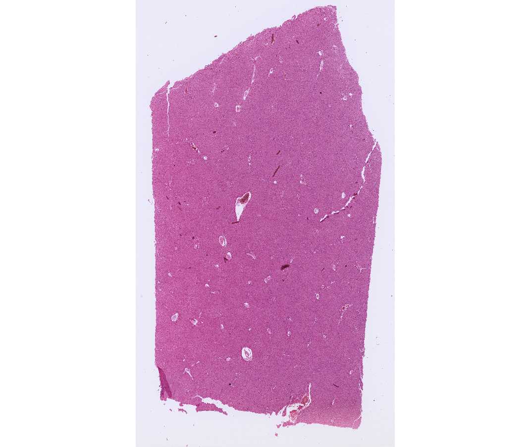
Identify the vessels and structures discussed above. Notice that a thin space is present between the endothelial cells lining the sinusoids and the parenchymal cells. This is the space of Disse, and it is in continuity with the lumen of the sinusoids via small spaces between the endothelial cells that form the wall of the sinusoids.
#47 Liver, Human (Phosphotungstic acid Hematoxylin - Phloxine)
Open with WebViewer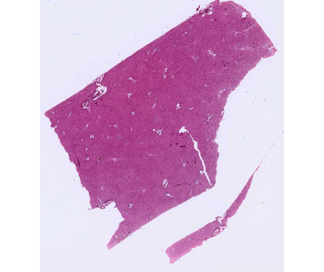
This slide should be examined as above. In addition, the bile canaliculi are revealed as delicate tubules that course between the apposed surfaces of the parenchymal cells. These are seen best in regions where the plates of cells are two cells thick.