SBPMD Histology Laboratory Manual
Gastrointestinal System II: Exocrine and Endocrine Pancreas
The pancreas develops as dorsal and ventral outgrowths of the duodenum, and its two diverticula fuse during later embryonic development. The pancreas contains both exocrine and endocrine components. The exocrine component consists of purely serous acini. It is the site of production and release of digestive enzymes (in an inactive state). These are delivered through a duct system that is similar to that in the salivary glands: intercalated duct to intralobular duct to interlobular duct. A diagnostic feature of the exocrine pancreas is the presence of centro-acinar cells. These cells form the initial portion of the intercalated duct. The pale-staining nuclei of the centro-acinar cells appear in the center of an acinus (hence their name).
Islets of Langerhans
The endocrine component of the pancreas consists of multiple spherical groups of epithelial cells embedded as nodules in the exocrine pancreas. The cells in these islets of Langerhans are not arranged into acini (as in the exocrine pancreas) but in irregular cords and clumps surrounded by a rich capillary plexus. Note that the islets are not separated from the acinar tissue by a capsule. The function of the islets is to control carbohydrate metabolism. The alpha cells secrete glucagon, which raises blood sugar, and the beta cells secrete insulin, which lowers it. There are several other biologically interesting peptides that are made by other cells of the islets. The most important identified to date, is somatostatin that is made by the delta cells. Its release locally inhibits both insulin and glucagon secretion.
#43 Pancreas, Guinea Pig (Chrome alum hematoxylin-phloxin) (not scanned)
Identify secretory acini and the duct system, which is particularly well defined in this preparation. The cytology of the acinar cells is best studied on this slide and slide #107. Note and understand the presence of intense cytoplasmic basophilia in the basal half of the pyramid-shaped acinar cells, and the accumulation of acidophilic granules in the apical cytoplasm.
The stain used in this slide allows you to distinguish cell types within the islets of Langerhans. Alpha cells stain red (phloxin) and beta cells stain blue (hematoxylin). The other cell types (e.g. delta cells) cannot be distinguished.
#42 Pancreas, Dog, H&E (not scanned)
Identify secretory acini and the duct system. The islets are pale as there is little cytoplasmic staining. You will not be able to distinguish alpha and beta cells on the basis of their staining properties in this H&E preparation.
#107 Pancreas thin section, Guinea pig. Acid fuchsin toluidine blue
Open with WebViewer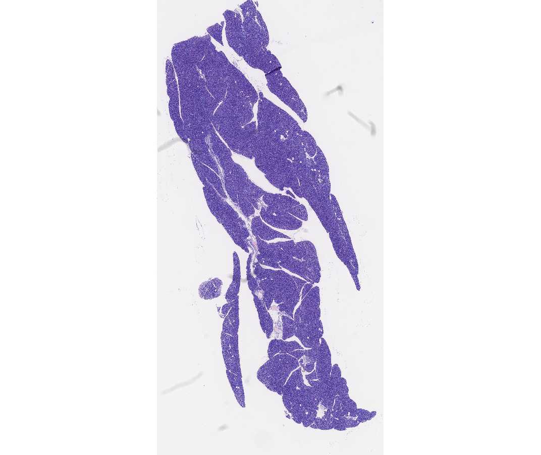
In the exocrine portion of the pancreas on this slide note that the basal cytoplasm of the acinar cells is highly basophilic. Zymogen granules at the apex are very acidophilic. The cytoplasm of centro-acinar cells and duct cells is relatively unstained. This is a good slide for studying the duct system. Islets of Langerhans are clearly visible, though again, the classes of hormone producing cells are not distinguishable.
Note: the following characteristics can be used to distinguish between the exocrine pancreas and the parotid. Islets of Langerhans are, of course, diagnostic for the pancreas, but they are not always seen in sections. In the pancreas, intralobular ducts are not conspicuous, and the centro-acinar cells are diagnostic. In contrast, acidophilic intralobular (striated) ducts are prominent in the parotid gland, and there are no centro-acinar cells.