SBPMD Histology Laboratory Manual
The Eye: Microscopic Examination
#119. Eye, sagittal section
Open with WebViewer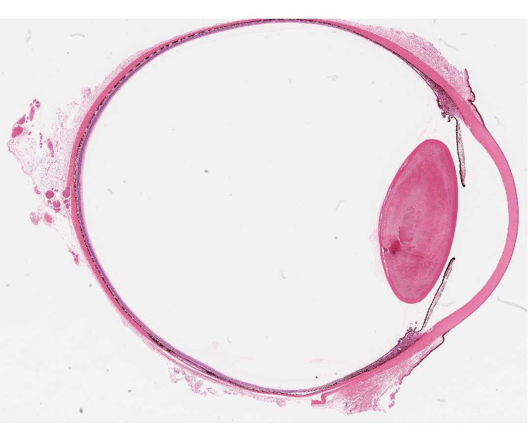
- Overview: The Layers Of The Eye: Corneosclera, Uvea, Retina
- The outermost layer: Corneosclera
- Cornea: The cornea is a transparent, avascular tissue composed of regularly arranged collagen fibrils and keratocytes (fibrocytes). Anteriorly it is lined with stratified squamous non-keratinized corneal epithelium with its underlying basal lamina and the acellular Bowman’s membrane. : Posteriorly it is lined by a single layer of flattened, hexagonal cells, the corneal endothelium that has a thick basal lamina, Descemet’s membrane.
- Conjunctiva: The conjunctiva is a thin, transparent mucous membrane that lines the posterior surfaces of the lids and the anterior part of the globe, up to the cornea.
- Sclera: The sclera is dense, irregularly arranged connective tissue, forming the white outer coat of the eye. The eye muscles insert on this coat.
- Limbus: The limbus is the transitional zone between the cornea and sclera. The region is referred to as the angle of the eye because in cross section a triangular space is formed between the cornea, sclera and ciliary body. In this region the aqueous humor leaves the anterior chamber via a trabecular meshwork, which merge to form the canal of Schlemm. The aqueous is then conveyed by "aqueous veins" to the blood vascular system in the sclera.
- The middle layer (uvea): Choroid, Iris and Ciliary Body
- Choroid: The choroid is a heavily vascularized and pigmented connective tissue whose boundary with the retinal pigment epithelium is Bruch's membrane (lamina vitrea), which is a thick basement membrane.
- Iris, with central aperture, the pupil: The iris projects from the choroid at the region of the limbus. It contains blood vessels, pigment cells, and muscles. Anteriorly it is a loose connective tissue containing pigment cells (melanocytes). The color of the iris (eye) depends on the number and disposition of the melanocytes in this layer. The anterior surface of the iris is an incomplete layer of fibroblasts. The deeper part of the connective tissue contains: blood vessels and nerves and the sphincter and dilator pupillae muscles. On the posterior side is a double layer of pigmented epithelial cells that are reflected at the pupillary margin. The basal lamina of the inner layer (posterior pigment epithelium) faces the lens. The basal lamina of the outer layer (anterior pigment epithelium) is adjacent to the stroma of the iris.
- Ciliary body:
At the base of the iris the ciliary body extends from the limbus (corneoscleral junction) to the ora serrata (the anterior limit of the retina and the choroid). Its outer side is adjacent to the sclera.
The ciliary body and its ciliary processes are covered by a two-layered epithelium: an outer pigmented ciliary epithelium that borders the connective tissue stroma and an inner non-pigmented ciliary epithelium that faces the posterior chamber. The ciliary body contains three groups of ciliary muscles embedded in the connective tissue. Two of these groups control the tension on the lens capsule and the other facilitates drainage of aqueous humor (discussed below). The ciliary processes are finger-like folds or outgrowths of the ciliary body to which the zonular fibers, which extend to the lens capsule are anchored and which contain large blood vessels and long fenestrated capillaries.
- The inner layer: Retina The two parts of the retina are the neural retina and the pigment epithelium. The neural retina, which contains photoreceptor cells that are responsible for absorbing light, has a laminated structure.
- Observation of the Contents of the Eye
- The anterior chamber, aqueous humor: The anterior chamber is the space between the cornea and the iris. It contains aqueous humor, which is produced by the ciliary processes. Aqueous humor passes from the posterior chamber into the anterior chamber between the iris and the lens. It is drained by the trabecular meshwork in the angle of the eye to the canal of Schlemm. This canal has openings that communicate via small aqueous veins with veins of the ciliary group.
- The posterior chamber, aqueous humor: The posterior chamber is the space between the posterior surface of the iris and the anterior surface of the lens.
- The lens and zonules: The lens is an avascular, non-innervated, epithelial tissue, which is bathed anteriorly by the aqueous humor and posteriorly by the vitreous. The anterior surface is covered by a monolayer of cuboidal epithelial cells. At the equator of the lens these cells are the progenitor cells for the lens fibers which comprise most of the lens mass. The lens is enveloped by a tremendously hypertrophied basal lamina called the lens capsule. The zonular fibers are inserted in the lens capsule and anchored in the basal lamina covering the ciliary epithelium. This arrangement provides the means of altering the tension on the zonules by contraction or relaxation of the ciliary muscle.
- Vitreous:
The vitreous is a loose connective tissue, which fills the center of the eye. The gel vitreous is made of thin, mostly randomly oriented, collagen filaments (type II collagen), which are anchored into the basal lamina of the retina, the ciliary body and the lens. There are no blood vessels or nerves in the vitreous, and there is only one layer of scattered phagocytic cells (hyalocytes) embedded in the outer margin of the gel in the vicinity of the retina and ciliary body.
- The Retina Identify the layers of the retina:
- Neural retina: The nuclei of the retinal cells form 3 layers: the outer nuclear layer, containing the nuclei of all photoreceptor cells; the inner nuclear layer, containing the nuclei of the bipolar, horizontal, amacrine, and Müller cells; and the ganglion cell layer containing the nuclei of the ganglion cells. The horizontal and amacrine cells are interneurons that integrate or associate the impulses from the photoreceptor and bipolar cells. The supporting framework of the retina is formed by the glial cells (Müller cells). Between the outer nuclear and inner nuclear layers is the outer plexiform layer. This contains the processes of rods and cones and of the bipolar cells and interneurons. Between the inner nuclear layer and the ganglion cell layer is the inner plexiform layer. This contains the processes of bipolar cells and interneurons and of ganglion cells. Internal to the ganglion cells the axons of these cells turn and course toward the optic disc, forming the nerve fiber layer. Internal to this is the basal lamina of the Müller cells, the internal limiting membrane.
- Retinal pigment epithelium: The inner surface of the retinal pigment epithelial cells has microvilli, which surround the outer segments of the photoreceptor cells. The retinal pigment epithelial cells also phagocytose packets of outer segment membrane, which are shed by the photoreceptor cells.
- Specialized regions of the retina (not present on your slides): The macula is a central area of the retina, about 5.5mm in diameter, having a high density of neurons (photoreceptor cells and the other retinal neurons). The center of the macula is the fovea, which provides us with high visual acuity, and which consists of densely packed cone photoreceptor cells. The inner layers of the retina are not present here. Ganglion cells are abundant around the fovea, supplying the foveal cones with a one to one connection and thus providing for maximal resolution. The optic disc is the area where all the ganglion cell axons exit. The main blood vessels of the retina enter and leave at the optic disc. It lacks all neural elements except for the axons of the ganglion cells. Therefore, it has no perceptive function and is the blind spot of the eye.